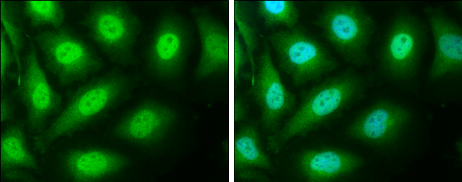
GTX112980 ICC/IF Image
SMAD4 antibody detects SMAD4 protein at cytoplasm and nucleus by immunofluorescent analysis.
Sample: HeLa cells were fixed in 4% paraformaldehyde at RT for 15 min.
Green: SMAD4 protein stained by SMAD4 antibody (GTX112980) diluted at 1:500.
Blue: Hoechst 33342 staining.
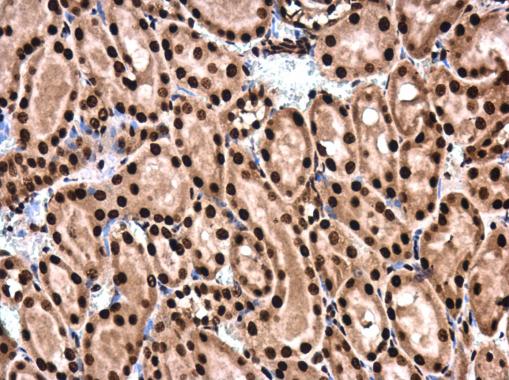
GTX112980 IHC-P Image
SMAD4 antibody detects SMAD4 protein at cytoplasm and nucleus in rat kidney by immunohistochemical analysis.
Sample: Paraffin-embedded rat kidney.
SMAD4 antibody (GTX112980) diluted at 1:500.
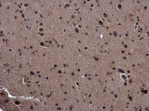
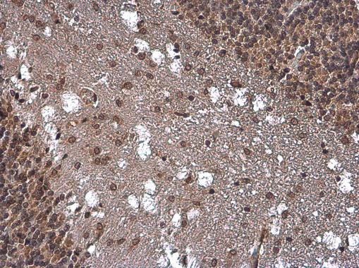
GTX112980 IHC-P Image
SMAD4 antibody detects SMAD4 protein at cytoplasm and nucleus in mouse brain by immunohistochemical analysis.
Sample: Paraffin-embedded mouse brain.
SMAD4 antibody (GTX112980) diluted at 1:500.
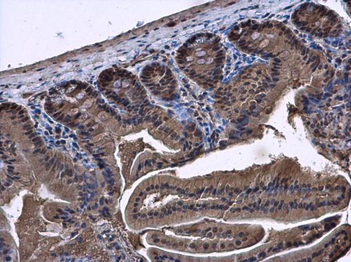
GTX112980 IHC-P Image
SMAD4 antibody detects SMAD4 protein at cytoplasm and nucleus in mouse duodenum by immunohistochemical analysis.
Sample: Paraffin-embedded mouse duodenum.
SMAD4 antibody (GTX112980) diluted at 1:500.
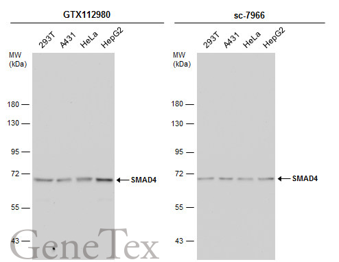
GTX112980 WB Image
Various whole cell extracts (30 ug) were separated by 7.5% SDS-PAGE, and the membranes were blotted with SMAD4 antibody (GTX112980) diluted at 1:1000 and competitor's antibody (sc-7966) diluted at 1:100. The HRP-conjugated anti-rabbit IgG antibody (GTX213110-01) was used to detect the primary antibody.
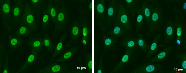
GTX112980 ICC/IF Image
SMAD4 antibody detects SMAD4 protein a tcytoplasm and nucleus by immunofluorescent analysis.
Sample: SK-N-SH cells were fixed in 4% paraformaldehyde at RT for 15 min.
Green: SMAD4 protein stained by SMAD4 antibody (GTX112980) diluted at 1:500.
Blue: Hoechst 33342 staining.
Scale bar = 10 um.
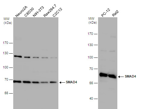
GTX112980 WB Image
SMAD4 antibody detects SMAD4 protein by western blot analysis. Various whole cell extracts were separated by 7.5% SDS-PAGE, and the membrane was blotted with SMAD4 antibody (GTX112980) diluted at 1:1000. The HRP-conjugated anti-rabbit IgG antibody (GTX213110-01) was used to detect the primary antibody.

GTX112980 IHC-P Image
Immunohistochemical analysis of paraffin-embedded human ulcerative colitis tissue using SMAD4 antibody (GTX112980)
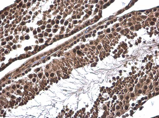
GTX112980 IHC-P Image
SMAD4 antibody detects SMAD4 protein at cytoplasm and nucleus by immunohistochemical analysis.Sample: Paraffin-embedded mouse testis.SMAD4 stained by SMAD4 antibody (GTX112980) diluted at 1:500.
GTX112980 IHC-P Image
SMAD4 antibody detects SMAD4 protein at cytoplasm and nucleus by immunohistochemical analysis.Sample: Paraffin-embedded rat brain.SMAD4 stained by SMAD4 antibody (GTX112980) diluted at 1:500.