GTX112883 ICC/IF Image
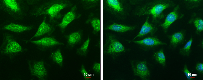
C1qA antibody detects C1qA protein at cytoplasm by immunofluorescent analysis.
Sample: HeLa cells were fixed in ice-cold MeOH for 5 min.
Green: C1qA protein stained by C1qA antibody (GTX112883) diluted at 1:100.
Blue: Hoechst 33342 staining.
Scale bar = 10 um.

GTX112883 WB Image
C1qA antibody detects C1QA protein by western blot analysis.
A. 50 ug mouse spleen lysate/extract
12% SDS-PAGE
C1qA antibody (GTX112883) dilution: 1:1000
The HRP-conjugated anti-rabbit IgG antibody (GTX213110-01) was used to detect the primary antibody.
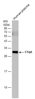
GTX112883 WB Image
Human tissue extract (30 ug) was separated by 12% SDS-PAGE, and the membrane was blotted with C1qA antibody (GTX112883) diluted at 1:1000. The HRP-conjugated anti-rabbit IgG antibody (GTX213110-01) was used to detect the primary antibody.