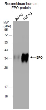
GTX112834 WB Image
Recombinant human EPO protein (with glycosylation) expressed in CHO cells (20 and 100 ng) were separated by 12% SDS-PAGE, and the membrane was blotted with (GTX112834) diluted at 1:20000.
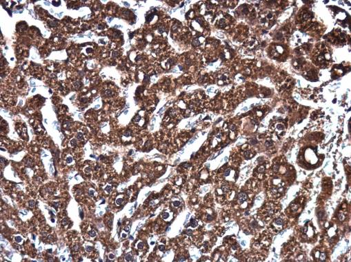
GTX112834 IHC-P Image
EPO antibody detects EPO protein at cytoplasm in rat liver by immunohistochemical analysis.
Sample: Paraffin-embedded rat liver.
EPO antibody (GTX112834) diluted at 1:400.
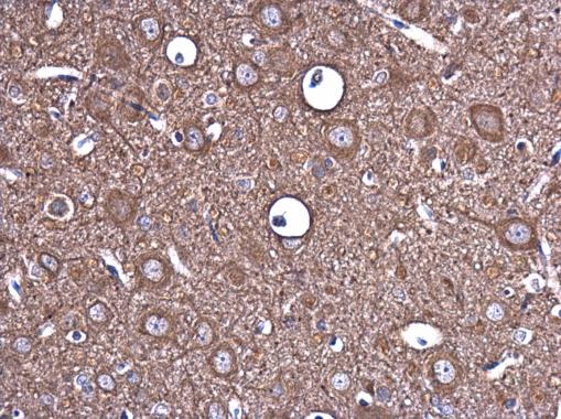
GTX112834 IHC-P Image
EPO antibody detects EPO protein at cytoplasm in rat brain by immunohistochemical analysis.
Sample: Paraffin-embedded rat brain.
EPO antibody (GTX112834) diluted at 1:400.
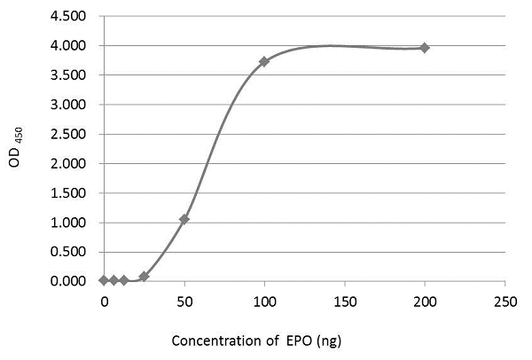
GTX112834 ELISA Image
An ELISA plate is coated with 50 uL of EPO recombinant protein at concentration ranged from 0.125 ug/mL to 4 ug/mL. The coated protein is detected with EPO antibody (GTX112834) at 1 ug/mL.