GTX112734 WB Image
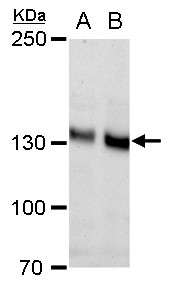
N-Cadherin antibody [N1N3] detects N-Cadherin protein by western blot analysis.
A. 30 ug PC-12 whole cell extract
B. 30 ug Rat2 whole cell extract
5% SDS-PAGE
N-Cadherin antibody [N1N3] (GTX112734) dilution: 1:1000
The HRP-conjugated anti-rabbit IgG antibody (GTX213110-01) was used to detect the primary antibody.
GTX112734 WB Image
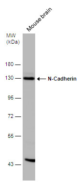
Mouse tissue extract (50 ug) was separated by 7.5% SDS-PAGE, and the membrane was blotted with N-Cadherin antibody [N1N3] (GTX112734) diluted at 1:500. The HRP-conjugated anti-rabbit IgG antibody (GTX213110-01) was used to detect the primary antibody.
GTX112734 IHC-P Image
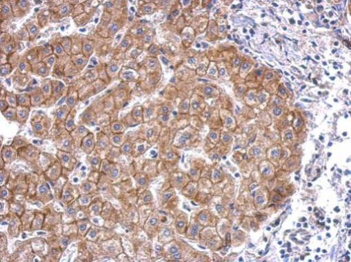
Immunohistochemical analysis of paraffin-embedded Mahlarvu xenograft, using N-Cadherin(GTX112734) antibody at 1:500 dilution.
GTX112734 IHC-P Image
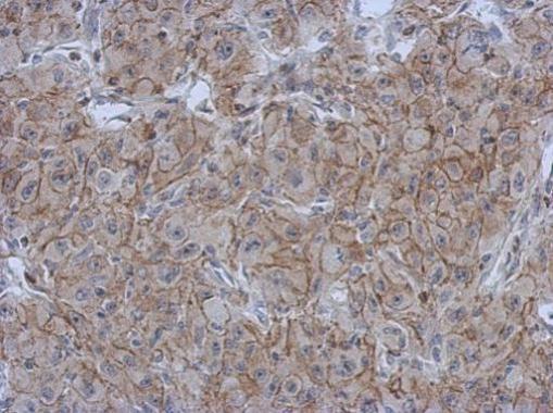
N-Cadherin antibody [N1N3] detects CDH2 protein at membrane on hepatoma by immunohistochemical analysis.
Sample: Paraffin-embedded human hepatoma.
N-Cadherin antibody [N1N3] (GTX112734) dilution: 1:500.
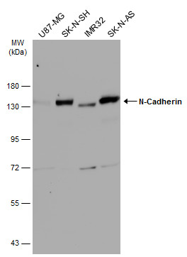
GTX112734 WB Image
Various whole cell extracts (30 ug) were separated by 7.5% SDS-PAGE, and the membrane was blotted with N-Cadherin antibody [N1N3] (GTX112734) diluted at 1:1000. The HRP-conjugated anti-rabbit IgG antibody (GTX213110-01) was used to detect the primary antibody.
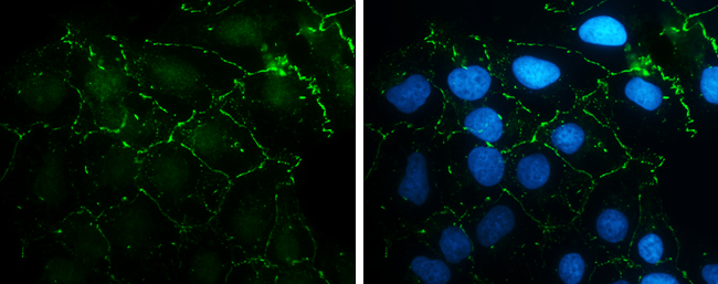
GTX112734 ICC/IF Image
N-Cadherin antibody [N1N3] detects N-Cadherin protein at cell membrane by immunofluorescent analysis.Sample: NT2D1 cells were fixed in 4% paraformaldehyde at RT for 15 min.Green: N-Cadherin stained by N-Cadherin antibody [N1N3] (GTX112734) diluted at 1:500.Blue: Hoechst 33342 staining.
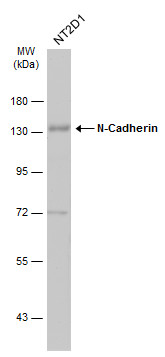
GTX112734 WB Image
Whole cell extract (30 ug) was separated by 7.5% SDS-PAGE, and the membrane was blotted with N-Cadherin antibody [N1N3] (GTX112734) diluted at 1:1000. The HRP-conjugated anti-rabbit IgG antibody (GTX213110-01) was used to detect the primary antibody.
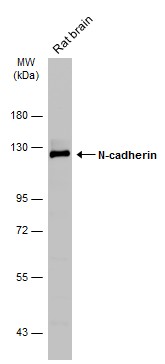
GTX112734 WB Image
Rat tissue extract (50 ug) was separated by 7.5% SDS-PAGE, and the membrane was blotted with N-cadherin antibody (GTX112734) diluted at 1:1000. The HRP-conjugated anti-rabbit IgG antibody (GTX213110-01) was used to detect the primary antibody.