GTX110587 ICC/IF Image
Immunofluorescence analysis of paraformaldehyde-fixed Human ESC, using STAT3(GTX110587) antibody at 1:200 dilution.
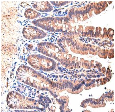
GTX110587 IHC-P Image
Immunohistochemical analysis of paraffin-embedded mouse small intestine, using STAT3(GTX110587) antibody at 1:200 dilution.(Image courtesy of Koji Taniguchi, Ph.D (Laboratory of Dr. Michael Karin, UCSD.)
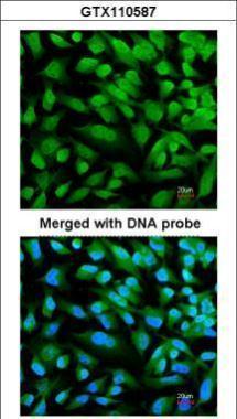
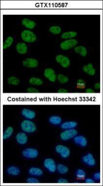
GTX110587 ICC/IF Image
Immunofluorescence analysis of paraformaldehyde-fixed HeLa, using STAT3(GTX110587) antibody at 1:200 dilution.
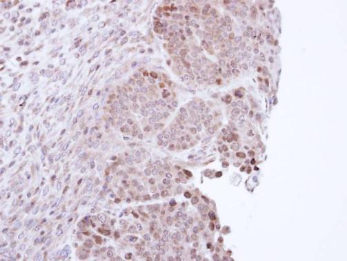
GTX110587 IHC-P Image
Immunohistochemical analysis of paraffin-embedded NCIN87 xenograft, using STAT3(GTX110587) antibody at 1:100 dilution.
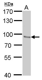
GTX110587 WB Image
STAT3 antibody detects STAT3 protein by western blot analysis.
A. 30 ug NIH-3T3 whole cell lysate/extract
7.5% SDS-PAGE
STAT3 antibody (GTX110587) dilution: 1:500
The HRP-conjugated anti-rabbit IgG antibody (GTX213110-01) was used to detect the primary antibody.
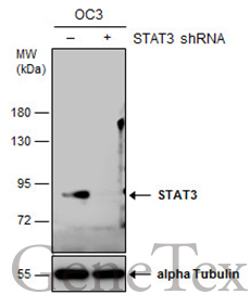
GTX110587 WB Image
Non-transfected (?) and transfected (+) OC3 whole cell extracts (30 ug) were separated by 7.5% SDS-PAGE, and the membrane was blotted with STAT3 antibody [C2C3], C-term (GTX110587) diluted at 1:500. The HRP-conjugated anti-rabbit IgG antibody (GTX213110-01) was used to detect the primary antibody.
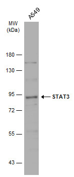
GTX110587 WB Image
Whole cell extract (30 ug) was separated by 7.5% SDS-PAGE, and the membrane was blotted with STAT3 antibody [C2C3], C-term (GTX110587) diluted at 1:500. The HRP-conjugated anti-rabbit IgG antibody (GTX213110-01) was used to detect the primary antibody.
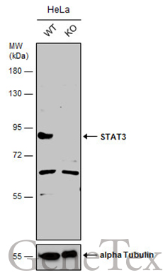
GTX110587 WB Image
Wild-type (WT) and STAT3 knockout (KO) HeLa cell extracts (30 ug) were separated by 7.5% SDS-PAGE, and the membrane was blotted with STAT3 antibody [C2C3], C-term (GTX110587) diluted at 1:500. The HRP-conjugated anti-rabbit IgG antibody (GTX213110-01) was used to detect the primary antibody, and the signal was developed with Trident ECL plus-Enhanced.