GTX110543 ICC/IF Image
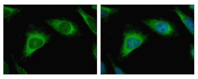
Caspase 3 antibody detects CASP3 protein at Cytoplasm by immunofluorescent analysis.
Sample: HeLa cells were fixed in 4% paraformaldehyde at RT for 15 min.
Green: CASP3 protein stained by Caspase 3 antibody (GTX110543) diluted at 1:500.
Blue: Hoechst 33343 staining.
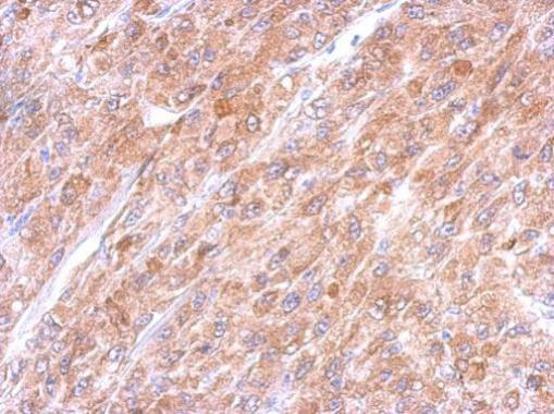
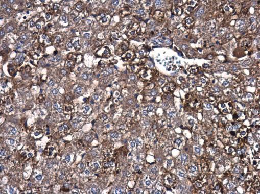
GTX110543 IHC-P Image
Immunohistochemical analysis of paraffin-embedded U87 xenograft, using Caspase 3(GTX110543) antibody at 1:500 dilution.
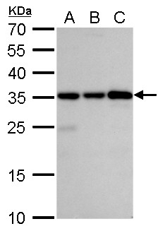
GTX110543 WB Image
Caspase 3 antibody detects Caspase 3 protein by western blot analysis.
A. 30 ug Jurkat whole cell lysate/extract
B. 30 ug Raji whole cell lysate/extract
C. 30 ug NCI-H929 whole cell lysate/extract
12% SDS-PAGE
Caspase 3 antibody (GTX110543) dilution: 1:5000
The HRP-conjugated anti-rabbit IgG antibody (GTX213110-01) was used to detect the primary antibody.
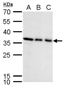
GTX110543 WB Image
Caspase 3 antibody detects Caspase 3 protein by western blot analysis.
A. 30 ug K562 whole cell lysate/extract
B. 30 ug THP-1 whole cell lysate/extract
C. 30 ug HL-60 whole cell lysate/extract
12% SDS-PAGE
Caspase 3 antibody (GTX110543) dilution: 1:5000
The HRP-conjugated anti-rabbit IgG antibody (GTX213110-01) was used to detect the primary antibody.
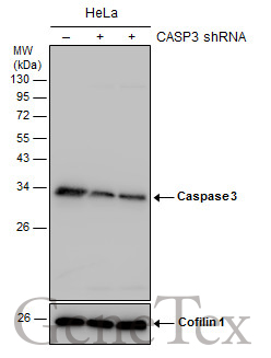
GTX110543 WB Image
Non-transfected (?) and transfected (+) HeLa whole cell extracts (30 ug) were separated by 12% SDS-PAGE, and the membrane was blotted with Caspase 3 antibody (GTX110543) diluted at 1:4000. The HRP-conjugated anti-rabbit IgG antibody (GTX213110-01) was used to detect the primary antibody.
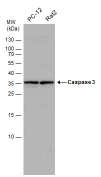
GTX110543 WB Image
Caspase 3 antibody detects Caspase 3 protein by western blot analysis. Various whole cell extracts (30 ug) were separated by 15% SDS-PAGE, and the membrane was blotted with Caspase 3 antibody (GTX110543) diluted by 1:1000. The HRP-conjugated anti-rabbit IgG antibody (GTX213110-01) was used to detect the primary antibody.
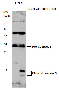
GTX110543 WB Image
Untreated (?) and treated (+) HeLa whole cell extracts (30 ug) were separated by 12% SDS-PAGE, and the membrane was blotted with Caspase 3 antibody (GTX110543) diluted at 1:1000. The HRP-conjugated anti-rabbit IgG antibody (GTX213110-01) was used to detect the primary antibody.
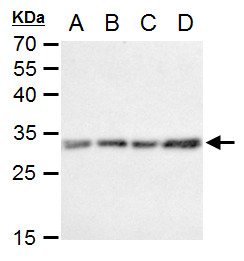
GTX110543 WB Image
Caspase 3 antibody detects Caspase 3 protein by western blot analysis.
A. 30 ug C8D30 whole cell extract
B. 30 ug NIH-3T3 whole cell extract
C. 30 ug Raw264.7 whole cell extract
D. 30 ug C2C12 whole cell extract
12% SDS-PAGE
Caspase 3 antibody (GTX110543) dilution: 1:1000
The HRP-conjugated anti-rabbit IgG antibody (GTX213110-01) was used to detect the primary antibody.
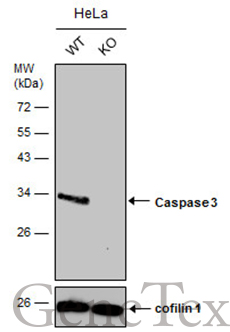
GTX110543 WB Image
Wild-type (WT) and Caspase 3 knockout (KO) HeLa cell extracts (30 ug) were separated by 12% SDS-PAGE, and the membrane was blotted with Caspase 3 antibody (GTX110543) diluted at 1:4000. The HRP-conjugated anti-rabbit IgG antibody (GTX213110-01) was used to detect the primary antibody.

GTX110543 WB Image
Caspase 3 antibody detects Caspase 3 protein by western blot analysis. Un-treated (-) and treated (+, 30 uM Cisplatin treatment for 24 hrs) PC-12 whole cell extracts (30 ug) were separated by 15% SDS-PAGE, and the membrane was blotted with Caspase 3 antibody (GTX110543) diluted by 1:1000. The HRP-conjugated anti-rabbit IgG antibody (GTX213110-01) was used to detect the primary antibody.
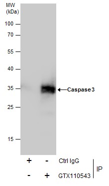
GTX110543 IP Image
Immunoprecipitation of Caspase 3 protein from HeLa whole cell extracts using 5 ug of Caspase 3 antibody (GTX110543).
Western blot analysis was performed using Caspase 3 antibody (GTX110543).
EasyBlot anti-Rabbit IgG (GTX221666-01) was used as a secondary reagent.
GTX110543 IHC-P Image
Caspase 3 antibody detects Caspase 3 protein at cytoplasm in rat liver by immunohistochemical analysis.
Sample: Paraffin-embedded rat liver.
Caspase 3 antibody (GTX110543) diluted at 1:500.