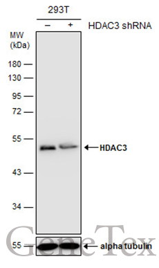
GTX109679 WB Image
Non-transfected (?) and transfected (+) 293T whole cell extracts (30 ug) were separated by 10% SDS-PAGE, and the membrane was blotted with HDAC3 antibody [C3], C-term (GTX109679) diluted at 1:500. The HRP-conjugated anti-rabbit IgG antibody (GTX213110-01) was used to detect the primary antibody.
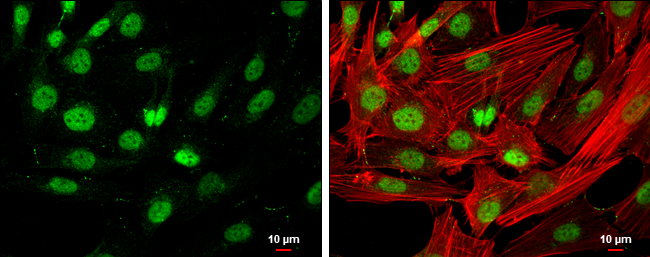
GTX109679 ICC/IF Image
HDAC3 antibody [C3], C-term detects HDAC3 protein at nucleus by immunofluorescent analysis.
Sample: SK-N-SH cells were fixed in 4% paraformaldehyde at RT for 15 min.
Green: HDAC3 protein stained by HDAC3 antibody [C3], C-term (GTX109679) diluted at 1:400.
Red: Phalloidin, a cytoskeleton marker, diluted at 1:200.
Scale bar = 10 um.
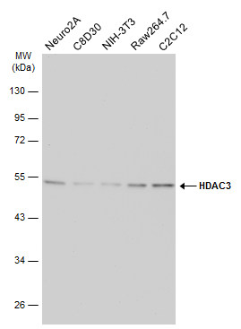
GTX109679 WB Image
Various whole cell extracts (30 ug) were separated by 10% SDS-PAGE, and the membrane was blotted with HDAC3 antibody [C3], C-term (GTX109679) diluted at 1:1000. The HRP-conjugated anti-rabbit IgG antibody (GTX213110-01) was used to detect the primary antibody.
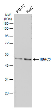
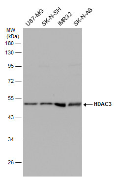
GTX109679 WB Image
Various whole cell extracts (30 ug) were separated by 10% SDS-PAGE, and the membrane was blotted with HDAC3 antibody [C3], C-term (GTX109679) diluted at 1:1000. The HRP-conjugated anti-rabbit IgG antibody (GTX213110-01) was used to detect the primary antibody.
GTX109679 WB Image
Various whole cell extracts (30 ug) were separated by 10% SDS-PAGE, and the membrane was blotted with HDAC3 antibody [C3], C-term (GTX109679) diluted at 1:500. The HRP-conjugated anti-rabbit IgG antibody (GTX213110-01) was used to detect the primary antibody.
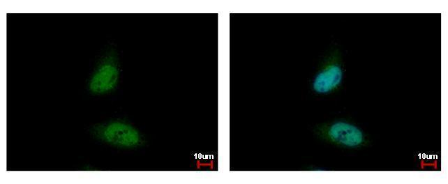
GTX109679 ICC/IF Image
HDAC3 antibody [C3], C-term detects HDAC3 protein at nucleus by immunofluorescent analysis.
Sample: HeLa cells were fixed in 4% paraformaldehyde at RT for 15 min.
Green: HDAC3 protein stained by HDAC3 antibody [C3], C-term (GTX109679) diluted at 1:500.
Blue: Hoechst 33342 staining.
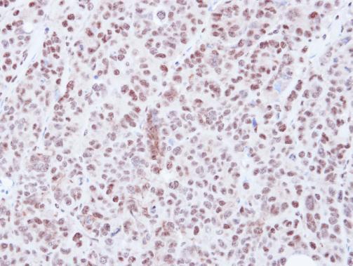
GTX109679 IHC-P Image
Immunohistochemical analysis of paraffin-embedded SW480 xenograft, using HDAC3(GTX109679) antibody at 1:500 dilution.
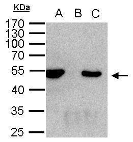
GTX109679 IP Image
HDAC3 antibody immunoprecipitates HDAC3 protein in IP experiments. IP Sample: 1000 ug 293T whole cell lysate/extract A. 50 ug 293T whole cell lysate/extract B. Control with 2 ug of preimmune rabbit IgG C. Immunoprecipitation of HDAC3 protein by 2 ug of HDAC3 antibody (GTX109679) 10% SDS-PAGE The immunoprecipitated HDAC3 protein was detected by HDAC3 antibody (GTX109679) diluted at 1:1000. EasyBlot anti-rabbit IgG (GTX221666-01) was used as a secondary reagent.
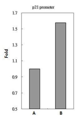
GTX109679 ChIP assay Image
HDAC3 antibody immunoprecipitates HDAC3 protein-DNA in ChIP experiments. ChIP Sample: 293T whole cell lysate/extract A. 5 ug preimmune rabbit IgG B. 5 ug of HDAC3 antibody (GTX109679) The precipitated DNA was detected by PCR with primer set targeting to p21 promoter.
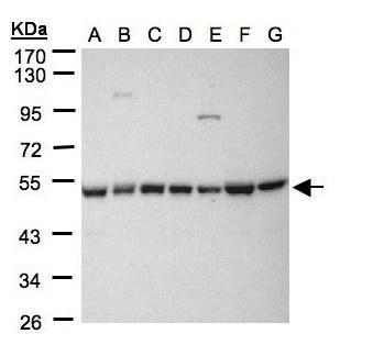
GTX109679 WB Image
Sample(30 ug whole cell lysate)
A: 293T
B: A431 (GTX27909)
C: H1299
D: HeLa S3 (GTX14654)
E: HepG2 (GTX27900)
F: MOLT4 (GTX27912)
G: Raji (GTX27908)
10% SDS PAGE
GTX109679 diluted at 1:1000
The HRP-conjugated anti-rabbit IgG antibody (GTX213110-01) was used to detect the primary antibody.