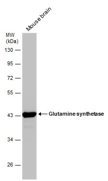
GTX109121 WB Image
Mouse tissue extract (50 ug) was separated by 10% SDS-PAGE, and the membrane was blotted with Glutamine synthetase antibody (GTX109121) diluted at 1:50000.
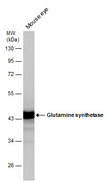
GTX109121 WB Image
Mouse tissue extract (50 ug) was separated by 10% SDS-PAGE, and the membrane was blotted with Glutamine synthetase antibody (GTX109121) diluted at 1:20000.
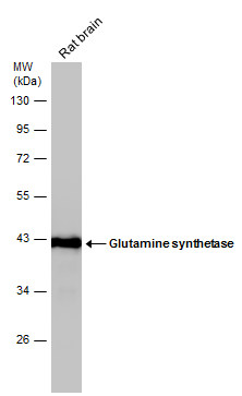
GTX109121 WB Image
Rat tissue extract (50 ug) was separated by 10% SDS-PAGE, and the membrane was blotted with Glutamine synthetase antibody (GTX109121) diluted at 1:50000.
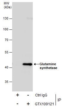
GTX109121 IP Image
Immunoprecipitation of Glutamine synthetase protein from IMR32 whole cell extracts using 5 ug of Glutamine synthetase antibody (GTX109121).
Western blot analysis was performed using Glutamine synthetase antibody (GTX109121).
EasyBlot anti-Rabbit IgG (GTX221666-01) was used as a secondary reagent.
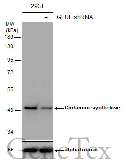
GTX109121 WB Image
Non-transfected (?) and transfected (+) 293T whole cell extracts (30 ug) were separated by 10% SDS-PAGE, and the membrane was blotted with Glutamine synthetase antibody (GTX109121) diluted at 1:1000. The HRP-conjugated anti-rabbit IgG antibody (GTX213110-01) was used to detect the primary antibody.
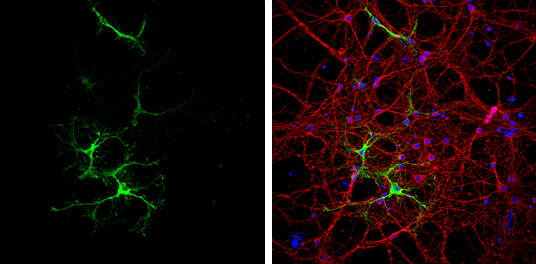
GTX109121 ICC/IF Image
Glutamine synthetase antibody detects Glutamine synthetase protein at astrocytes by immunofluorescent analysis.
Sample: DIV9 rat E18 primary cortical neurons were fixed in 4% paraformaldehyde at RT for 15 min.
Green: Glutamine synthetase protein stained by Glutamine synthetase antibody (GTX109121) diluted at 1:500.
Red: beta Tubulin 3/ Tuj1, a neuron cell marker, stained by beta Tubulin 3/ Tuj1 antibody [GT11710] (GTX631836) diluted at 1:500.
Blue: Fluoroshield with DAPI (GTX30920).
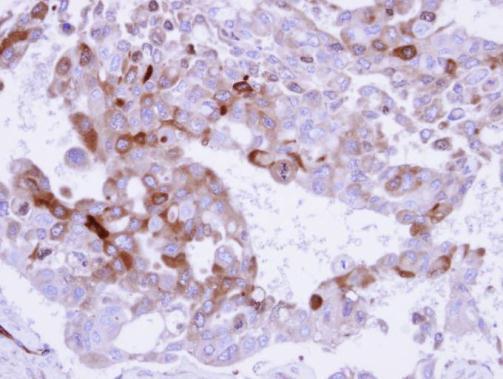
GTX109121 IHC-P Image
Immunohistochemical analysis of paraffin-embedded H441 xenograft , using Glutamine Synthetase (GTX109121) antibody at 1:500 dilution.
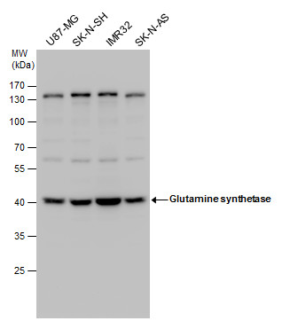
GTX109121 WB Image
Glutamine synthetase antibody detects Glutamine synthetase protein by western blot analysis. Various whole cell extracts (30 ug) were separated by 10% SDS-PAGE, and the membrane was blotted with Glutamine synthetase antibody (GTX109121) diluted at a dilution of 1:5000. The HRP-conjugated anti-rabbit IgG antibody (GTX213110-01) was used to detect the primary antibody.
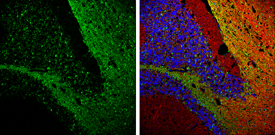
GTX109121 IHC-Fr Image
Glutamine synthetase antibody detects Glutamine synthetase protein expression by immunohistochemical analysis.
Sample: Frozen-sectioned adult mouse cerebellum.
Green: Glutamine synthetase protein stained by Glutamine synthetase antibody (GTX109121) diluted at 1:250.
Red: beta Tubulin 3/ TUJ1, stained by beta Tubulin 3/ TUJ1 antibody [GT11710] (GTX631836) diluted at 1:500.
Blue: Fluoroshield with DAPI (GTX30920).
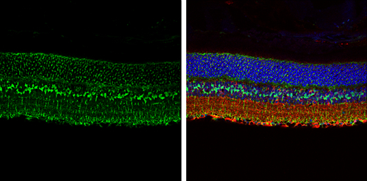
GTX109121 IHC-P Image
Glutamine synthetase antibody detects Glutamine synthetase protein expression by immunohistochemical analysis.
Sample:Paraffin-embedded adult mouse retina.
Green: Glutamine synthetase protein stained by Glutamine synthetase antibody (GTX109121) diluted at 1:250.
Red: beta Tubulin 3/ TUJ1, stained by beta Tubulin 3/ TUJ1 antibody [GT11710] (GTX631836) diluted at 1:250.
Blue: Fluoroshield with DAPI (GTX30920).