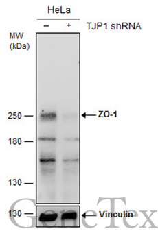
GTX108627 WB Image
Non-transfected (?) and transfected (+) HeLa whole cell extracts (30 ug) were separated by 5% SDS-PAGE, and the membrane was blotted with ZO-1 antibody [N2C1], Internal (GTX108627) diluted at 1:500. The HRP-conjugated anti-rabbit IgG antibody (GTX213110-01) was used to detect the primary antibody.
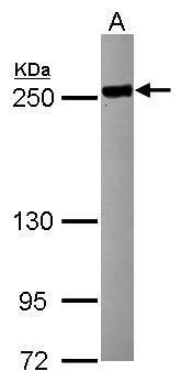
GTX108627 WB Image
Sample (30 ug of whole cell lysate)
A: HCT116
5% SDS PAGE
GTX108627 diluted at 1:5000
The HRP-conjugated anti-rabbit IgG antibody (GTX213110-01) was used to detect the primary antibody.
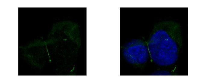
GTX108627 ICC/IF Image
ZO-1 antibody [N2C1], Internal detects TJP1 protein at junction by confocal immunofluorescent analysis.
Sample: A431 cells were fixed in ice-cold MeOH for 5 min.
Green: TJP1 protein stained by ZO-1 antibody [N2C1], Internal (GTX108627) diluted at 1:500.
Blue: Hoechst 33342 staining.
[Images captured by Olympus FV10i Confocal Laser Scanning Microscope]