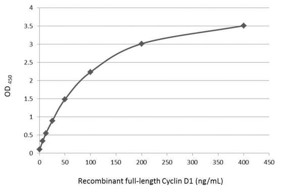
GTX108624 ELISA Image
Sandwich ELISA detection of recombinant full-length Cyclin D1 protein using GTX634347 as capture antibody at concentration of 5 ug/mL and GTX108624 as detection antibody at concentration of 1 ug/mL. Rabbit IgG antibody (HRP) (GTX213110-01) was diluted at 1:10000 and used to detect the primary antibody.
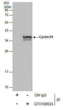
GTX108624 IP Image
Immunoprecipitation of Cyclin D1 protein from MCF-7 whole cell extracts using 5 ug of Cyclin D1 antibody [N1C3] (GTX108624).
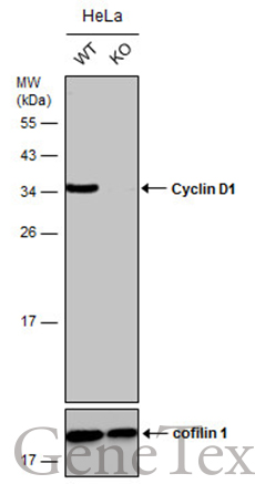
Western blot analysis was performed using Cyclin D1 antibody [N1C3] (GTX108624).
EasyBlot anti-Rabbit IgG (GTX221666-01) was used as a secondary reagent.
GTX108624 WB Image
Wild-type (WT) and Cyclin D1 knockout (KO) HeLa cell extracts (30 ug) were separated by 12% SDS-PAGE, and the membrane was blotted with Cyclin D1 antibody [N1C3] (GTX108624) diluted at 1:5000. The HRP-conjugated anti-rabbit IgG antibody (GTX213110-01) was used to detect the primary antibody, and the signal was developed with Trident ECL plus-Enhanced.
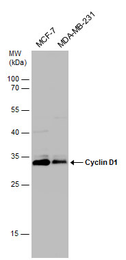
GTX108624 WB Image
Various whole cell extracts (30 ug) were separated by 12% SDS-PAGE, and the membrane was blotted with Cyclin D1 antibody [N1C3] (GTX108624) diluted at 1:1000.
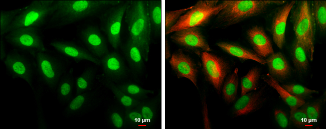
GTX108624 ICC/IF Image
Cyclin D1 antibody [N1C3] detects Cyclin D1 protein at cytoplasm and nucleus by immunofluorescent analysis.
Sample: SK-N-SH cells were fixed in 4% paraformaldehyde at RT for 15 min.
Green: Cyclin D1 protein stained by Cyclin D1 antibody [N1C3] (GTX108624) diluted at 1:500.
Red: beta Tubulin 3/ Tuj1, a cytoskeleton marker, stained by beta Tubulin 3/ Tuj1 antibody [GT11710] (GTX631836) diluted at 1:500.
Scale bar = 10 um.