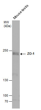
GTX108613 WB Image
Mouse tissue extract (50 ug) was separated by 5% SDS-PAGE, and the membrane was blotted with ZO-1 antibody [N1N2], N-term (GTX108613) diluted at 1:500. The HRP-conjugated anti-rabbit IgG antibody (GTX213110-01) was used to detect the primary antibody.
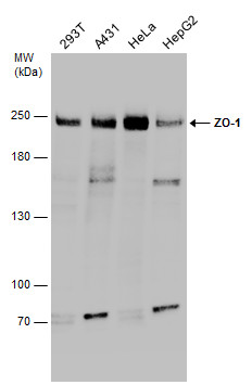
GTX108613 WB Image
Various whole cell extracts (30 ug) were separated by 5% SDS-PAGE, and the membrane was blotted with ZO-1 antibody [N1N2], N-term (GTX108613) diluted at 1:1000.
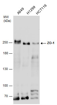
GTX108613 WB Image
Various whole cell extracts (30 ug) were separated by 5% SDS-PAGE, and the membrane was blotted with ZO-1 antibody [N1N2], N-term (GTX108613) diluted at 1:3000.
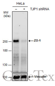
GTX108613 WB Image
Non-transfected (?) and transfected (+) HeLa whole cell extracts (30 ug) were separated by 5% SDS-PAGE, and the membrane was blotted with ZO-1 antibody [N1N2], N-term (GTX108613) diluted at 1:1000. The HRP-conjugated anti-rabbit IgG antibody (GTX213110-01) was used to detect the primary antibody.
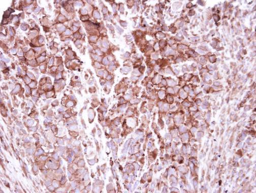
GTX108613 IHC-P Image
Immunohistochemical analysis of paraffin-embedded H661 xenograft, using ZO-1(GTX108613) antibody at 1:250 dilution.
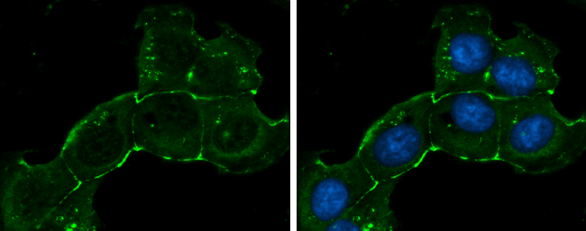
GTX108613 ICC/IF Image
ZO-1 antibody [N1N2], N-term detects ZO-1 protein at cell membrane by immunofluorescent analysis.Sample: NT2D1 cells were fixed in ice-cold MeOH for 5 min.Green: ZO-1 stained by ZO-1 antibody [N1N2], N-term (GTX108613) diluted at 1:200.Blue: Hoechst 33342 staining.
GTX108613 IHC-P Image
ZO-1 antibody [N1N2], N-term detects ZO-1 protein at cell membrane and cytoplasm in mouse testis by immunohistochemical analysis.
Sample: Paraffin-embedded mouse testis.
ZO-1 antibody [N1N2], N-term (GTX108613) diluted at 1:500.

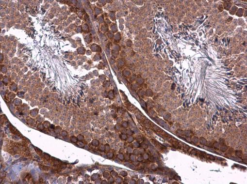
GTX108613 IHC-P Image
ZO-1 antibody [N1N2], N-term detects ZO-1 protein at cell membrane and cytoplasm in mouse intestine by immunohistochemical analysis.
Sample: Paraffin-embedded mouse intestine.
ZO-1 antibody [N1N2], N-term (GTX108613) diluted at 1:500.