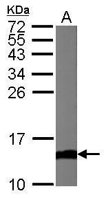
GTX108585 WB Image
Sample (50 ug of whole cell lysate)
A: Mouse brain
15% SDS PAGE
GTX108585 diluted at 1:1000
The HRP-conjugated anti-rabbit IgG antibody (GTX213110-01) was used to detect the primary antibody.
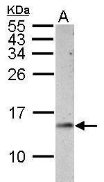
GTX108585 WB Image
Sample (30 ug of whole cell lysate)
A: PC-12
15% SDS PAGE
GTX108585 diluted at 1:1000
The HRP-conjugated anti-rabbit IgG antibody (GTX213110-01) was used to detect the primary antibody.
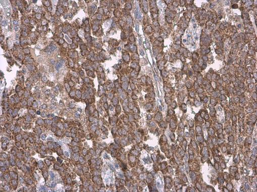
GTX108585 IHC-P Image
Cytochrome C antibody detects Cytochrome C protein at cytoplasm by immunohistochemical analysis.Sample: Paraffin-embedded human endometrial carcinoma.Cytochrome C stained by Cytochrome C antibody (GTX108585) diluted at 1:500.
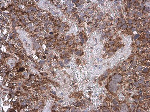
GTX108585 IHC-P Image
Cytochrome C antibody detects Cytochrome C protein at cytoplasm by immunohistochemical analysis.Sample: Paraffin-embedded human cervical carcinoma.Cytochrome C stained by Cytochrome C antibody (GTX108585) diluted at 1:500.
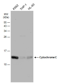
GTX108585 WB Image
Various whole cell extracts (30 ug) were separated by 15% SDS-PAGE, and the membrane was blotted with Cytochrome C antibody (GTX108585) diluted at 1:1000. The HRP-conjugated anti-rabbit IgG antibody (GTX213110-01) was used to detect the primary antibody.