
GTX107562 ICC/IF Image
Confocal immunofluorescence analysis (Olympus FV10i) of paraformaldehyde-fixed HeLa, using p70 S6K(GTX107562) antibody (Green) at 1:500 dilution. Alpha-tubulin filaments were labeled with GTX11304 (Red) at 1:2000.

GTX107562 IHC-P Image
Immunohistochemical analysis of paraffin-embedded SW480 xenograft, using p70 S6K(GTX107562) antibody at 1:100 dilution.
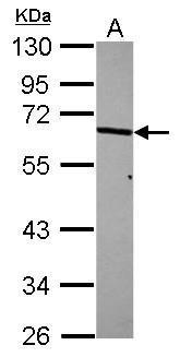
GTX107562 WB Image
Sample (30 ug of whole cell lysate)
A: A431
10% SDS PAGE
GTX107562 diluted at 1:1000
The HRP-conjugated anti-rabbit IgG antibody (GTX213110-01) was used to detect the primary antibody.

GTX107562 IHC-P Image
p70 S6K antibody detects RPS6KB1 protein at cytoplasm in rat kidney by immunohistochemical analysis.
Sample: Paraffin-embedded rat kidney.
p70 S6K antibody (GTX107562) diluted at 1:500.

GTX107562 WB Image
Various whole cell extracts (30 ug) were separated by 10% SDS-PAGE, and the membrane was blotted with p70 S6K antibody (GTX107562) diluted at 1:1000. The HRP-conjugated anti-rabbit IgG antibody (GTX213110-01) was used to detect the primary antibody.
GTX107562 WB Image
Whole cell extract (30 ug) was separated by 10% SDS-PAGE, and the membrane was blotted with p70 S6K antibody (GTX107562) diluted at 1:1000. The HRP-conjugated anti-rabbit IgG antibody (GTX213110-01) was used to detect the primary antibody.

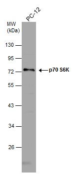
GTX107562 WB Image
S6K antibody detects S6K protein by western blot analysis. Whole cell extracts (30 ug) was separated by 10% SDS-PAGE, and the membrane was blotted with S6K antibody (GTX107562) at a dilution of 1:1000. The HRP-conjugated anti-rabbit IgG antibody (GTX213110-01) was used to detect the primary antibody.