GTX10549 WB Image
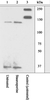
Lysates prepared from MDCK cells left untreated (1, 3) or treated with staurosporine (2) were resolved by SDS-PAGE on a 10% polyacrylamide gel and transferred to PVDF. Membranes were blocked with a 5% BSA-TBST buffer for one hour at room temperature, and incubated with ROCK1 (GTX10549) cleavage-site specific antibody (1, 2) or a ROCK1 pan antibody (3) in a 3% BSA-TBST buffer for two hours at room temperature. After washing, membranes were incubated with goat F(abÅf)2 anti-rabbit IgG HRP conjugate and bands were detected. The data show that ROCK1 [1113/1114] CSSA detected the 130 kDa cleaved form of ROCK but not the full length. This antibody did not detect the 130 kDa cleaved form of ROCK upon inhibition of caspase activity with the pan caspase inhibitor Ac-VAD-CHO (data not shown).