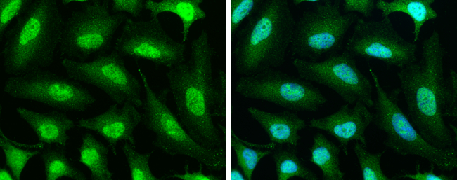
GTX105295 ICC/IF Image
PPP1CB antibody detects PPP1CB protein at cytoplasm and nucleus by immunofluorescent analysis.Sample: HeLa cells were fixed in 4% paraformaldehyde at RT for 15 min.Green: PPP1CB stained by PPP1CB antibody (GTX105295) diluted at 1:500.Blue: Hoechst 33342 staining.
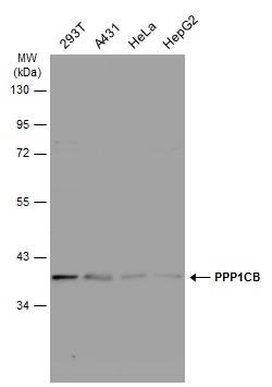
GTX105295 WB Image
Various whole cell extracts (30 ug) were separated by 10% SDS-PAGE, and the membrane was blotted with PPP1CB antibody (GTX105295) diluted at 1:1000. The HRP-conjugated anti-rabbit IgG antibody (GTX213110-01) was used to detect the primary antibody.
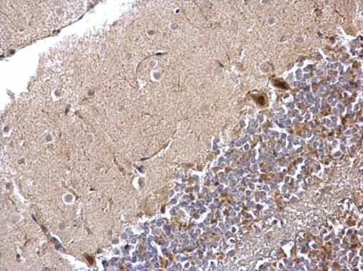
GTX105295 IHC-P Image
PPP1CB antibody detects PPP1CB protein at nucleus and cytosol on mouse hind brain by immunohistochemical analysis.
Sample: Paraffin-embedded mouse hind brain.
PPP1CB antibody (GTX105295) dilution: 1:500.
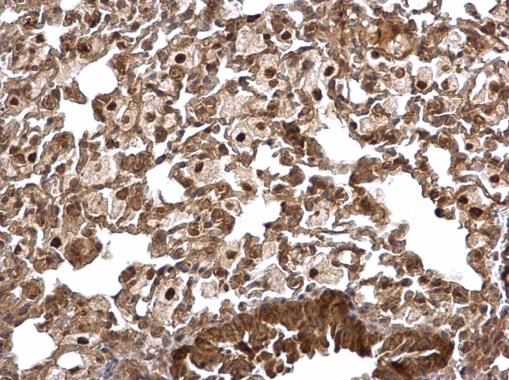
GTX105295 IHC-P Image
PPP1CB antibody detects PPP1CB protein at nucleus and cytosol on mouse lung by immunohistochemical analysis.
Sample: Paraffin-embedded mouse lung.
PPP1CB antibody (GTX105295) dilution: 1:500.
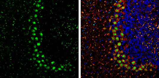
GTX105295 IHC-Fr Image
PPP1CB antibody detects PPP1CB Protein expression by immunohistochemical analysis.
Sample: Frozen-sectioned adult mouse cerebellum.
Green: PPP1CB stained by PPP1CB antibody (GTX105295) diluted at 1:250.
Red: NF-H, stained by NF-H antibody [GT114] (GTX634289) diluted at 1:500.
Blue: Fluoroshield with DAPI (GTX30920).
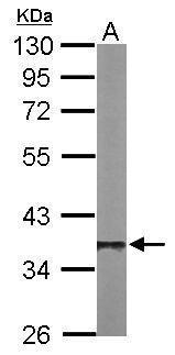
GTX105295 WB Image
Sample (50 ug of whole cell lysate)
A: Mouse brain
10% SDS PAGE
GTX105295 diluted at 1:1000
The HRP-conjugated anti-rabbit IgG antibody (GTX213110-01) was used to detect the primary antibody.