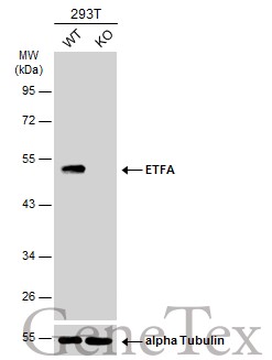
GTX105155 WB Image
Wild-type (WT) and ETFA knockout (KO) 293T cell extracts (30 ug) were separated by 12% SDS-PAGE, and the membrane was blotted with ETFA antibody (GTX105155) diluted at 1:1000. The HRP-conjugated anti-rabbit IgG antibody (GTX213110-01) was used to detect the primary antibody.
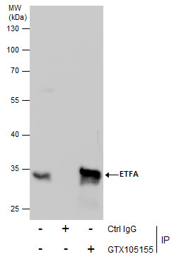
GTX105155 IP Image
Immunoprecipitation of ETFA protein from HepG2 whole cell extracts using 5 ug of ETFA antibody (GTX105155).
Western blot analysis was performed using ETFA antibody (GTX105155).
EasyBlot anti-Rabbit IgG (GTX221666-01) was used as a secondary reagent.
GTX105155 IHC-P Image
ETFA antibody detects ETFA protein at mitochondria on mouse liver by immunohistochemical analysis.
Sample: Paraffin-embedded mouse heart.
ETFA antibody (GTX105155) dilution: 1:500.
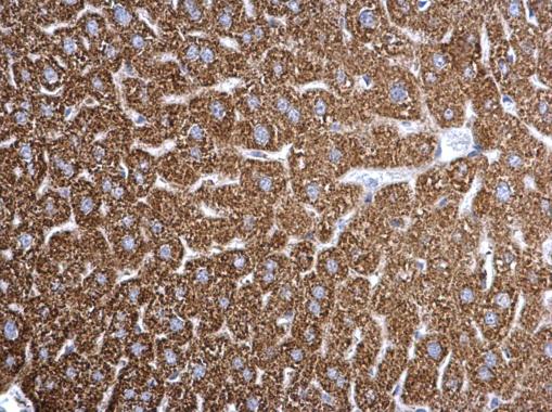
GTX105155 IHC-P Image
ETFA antibody detects ETFA protein at mitochondria on mouse liver by immunohistochemical analysis.
Sample: Paraffin-embedded mouse liver.
ETFA antibody (GTX105155) dilution: 1:500.
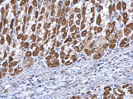
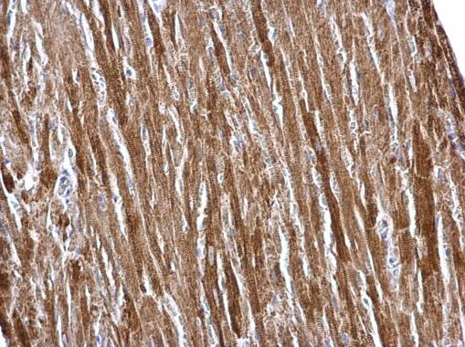
GTX105155 IHC-P Image
ETFA antibody detects ETFA protein at mitochondria on mouse heart by immunohistochemical analysis.
Sample: Paraffin-embedded mouse stomach.
ETFA antibody (GTX105155) dilution: 1:500.
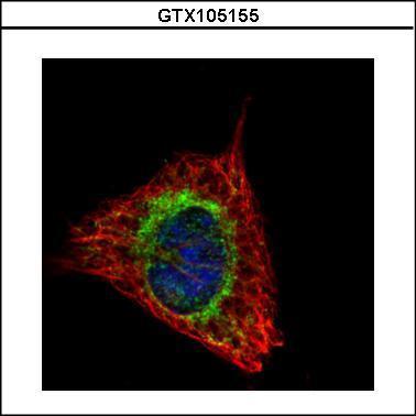
GTX105155 ICC/IF Image
Confocal immunofluorescence analysis (Olympus FV10i) of methanol-fixed HeLa, using ETFA(GTX105155) antibody (Green) at 1:500 dilution. Alpha-tubulin filaments were labeled with GTX11304 (Red) at 1:2000.
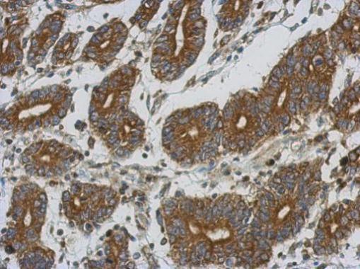
GTX105155 IHC-P Image
Immunohistochemical analysis of paraffin-embedded human colon carcinoma, using ETFA(GTX105155) antibody at 1:500 dilution.
GTX105155 WB Image
Sample (30 ug of whole cell lysate)
A: A431
B: HeLa
10% SDS PAGE
GTX105155 diluted at 1:1000
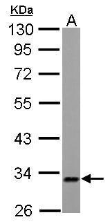
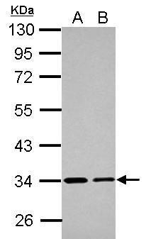
GTX105155 WB Image
ETFA antibody detects ETFA protein by Western blot analysis.
A. 30 ug PC-12 whole cell lysate/extract
10 % SDS-PAGE
ETFA antibody (GTX105155) dilution: 1:1000