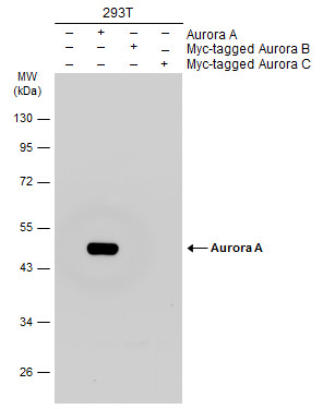
GTX104620 WB Image
Non-transfected (?) and transfected (+) 293T whole cell extracts (30 ug) were separated by 10% SDS-PAGE, and the membrane was blotted with Aurora A antibody [C3], C-term (GTX104620) diluted at 1:1000. The HRP-conjugated anti-rabbit IgG antibody (GTX213110-01) was used to detect the primary antibody.
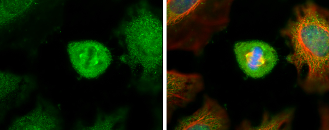
GTX104620 ICC/IF Image
Aurora A antibody [C3], C-term detects Aurora A protein at spindle by immunofluorescent analysis.Sample: HeLa cells were fixed in ice-cold MeOH for 5 min.Green: Aurora A stained by Aurora A antibody [C3], C-term (GTX104620) diluted at 1:500.Red: alpha Tubulin, a cytoskeleton marker, stained by alpha Tubulin antibody [GT114] (GTX628802) diluted at 1:500.Blue: Hoechst 33342 staining.
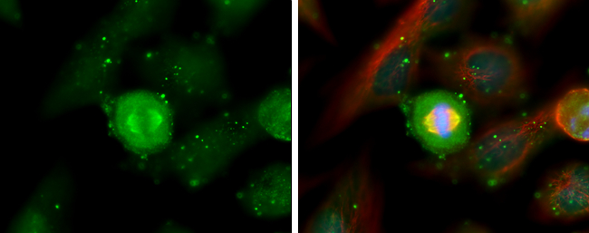
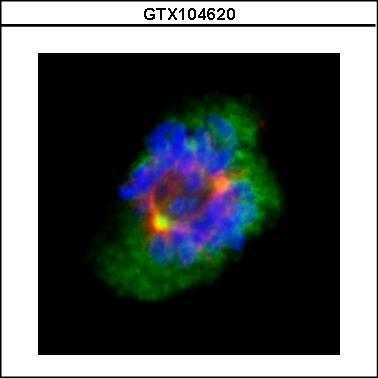
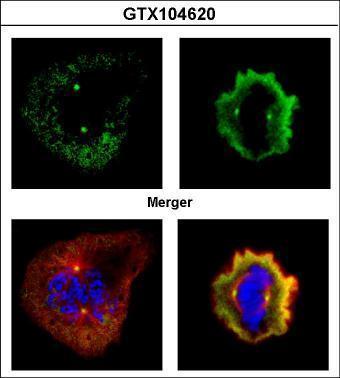
GTX104620 ICC/IF Image
Confocal immunofluorescence analysis (Olympus FV10i) of methanol-fixed 293T, using Aurora A(GTX104620) antibody (Green) at 1:500 dilution. Alpha-tubulin filaments were labeled with GTX11304 (Red) at 1:2500.
GTX104620 ICC/IF Image
Confocal immunofluorescence analysis (Olympus FV10i) of paraformaldehyde-fixed U2OS, using Aurora A(GTX104620) antibody (Green) at 1:500 dilution. Alpha-tubulin filaments were labeled with GTX11304 (Red) at 1:500.
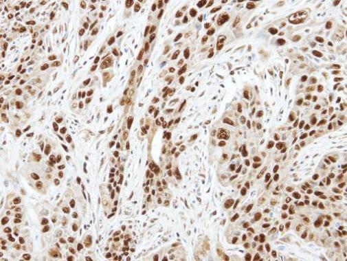
GTX104620 IHC-P Image
Immunohistochemical analysis of paraffin-embedded TW2.6 xenograft, using Aurora A(GTX104620) antibody at 1:100 dilution.