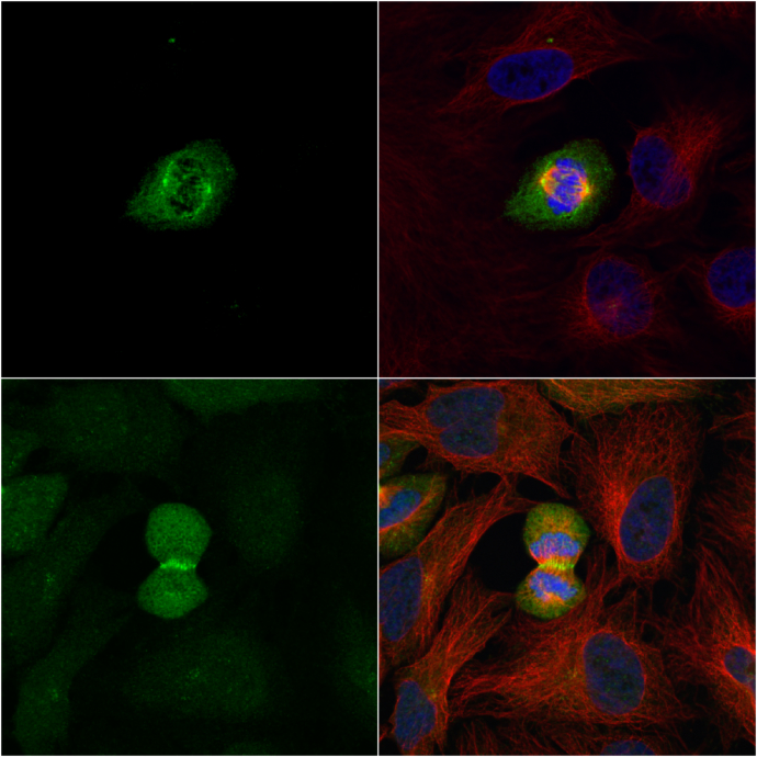
GTX104302 ICC/IF Image
PLK1 antibody [N2C2], Internal detects PLK1 protein at midbody by immunofluorescent analysis.
Sample: HeLa cells were fixed in 2% paraformadehyde at RT for 30 min.
Green: PLK1 protein stained by PLK1 antibody [N2C2], Internal (GTX104302) diluted at 1:1000.
Red: alpha Tubulin, a cytoskeleton marker, stained by alpha Tubulin antibody [GT114] (GTX628802) diluted at 1:1000.
Blue: Hoechst 33342 staining.
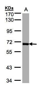
GTX104302 WB Image
Sample(30 ug whole cell lysate)
A:Raji (GTX27908)
7.5% SDS PAGE
GTX104302 diluted at 1:1000
The HRP-conjugated anti-rabbit IgG antibody (GTX213110-01) was used to detect the primary antibody.

GTX104302 IHC-P Image
Immunohistochemical analysis of paraffin-embedded human skin, using PLK1(GTX104302) antibody(10 ug/ml).