
GTX104126 WB Image
Various whole cell extracts (30 ug) were separated by 7.5% SDS-PAGE, and the membrane was blotted with HSPA1A antibody (GTX104126) diluted at 1:30000. The HRP-conjugated anti-rabbit IgG antibody (GTX213110-01) was used to detect the primary antibody.
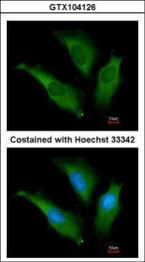
GTX104126 ICC/IF Image
Immunofluorescence analysis of methanol-fixed HeLa, using HSP70 1A(GTX104126) antibody at 1:200 dilution.
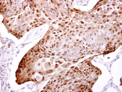
GTX104126 IHC-P Image
HSP70 1A antibody detects HSP70 1A protein at cytosol and nucleus on human breast carcinoma by immunohistochemical analysis.
Sample: Paraffin-embedded human breast carcinoma.
HSP70 1A antibody (GTX104126) dilution: 1:500.

GTX104126 WB Image
Sample (50 ug of whole cell lysate)
A: Mouse brain
7.5% SDS PAGE
GTX104126 diluted at 1:1000
The HRP-conjugated anti-rabbit IgG antibody (GTX213110-01) was used to detect the primary antibody.

GTX104126 IP Image
HSP70 1A antibody immunoprecipitates HSP70 1A protein in IP experiments. IP Sample: 1000 ug HeLa whole cell lysate/extract A. 40 ug HeLa whole cell lysate/extract B. Control with 2.5 ug of preimmune rabbit IgG C. Immunoprecipitation of HSP70 1A protein by 2.5 ug of HSP70 1A antibody (GTX104126) 12% SDS-PAGE The immunoprecipitated HSP70 1A protein was detected by HSP70 1A antibody (GTX104126) diluted at 1:1000. EasyBlot anti-rabbit IgG (GTX221666-01) was used as a secondary reagent.
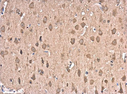
GTX104126 IHC-P Image
HSP70 1A antibody detects HSP70 1A protein at cytoplasm in rat brain by immunohistochemical analysis.
Sample: Paraffin-embedded rat brain.
HSP70 1A antibody (GTX104126) diluted at 1:500.

GTX104126 WB Image
Rat tissue extract (50 ug) was separated by 7.5% SDS-PAGE, and the membrane was blotted with HSPA1A antibody (GTX104126) diluted at 1:1000.