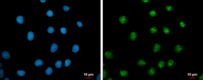
GTX103436 ICC/IF Image
c-Myc antibody detects c-Myc protein at nucleus by immunofluorescent analysis.
Sample: NT2D1 cells were fixed in 4% paraformaldehyde at RT for 15 min.
Green: c-Myc protein stained by c-Myc antibody (GTX103436) diluted at 1:200.
Blue: Hoechst 33342 staining.
Scale bar = 10 um.
GTX103436 IP Image
c-Myc antibody immunoprecipitates c-Myc protein in IP experiments. IP Sample: 1000 ug 293T whole cell lysate/extract A. 20 ug 293T whole cell lysate/extract B. Control with 2.5 ug of preimmune rabbit IgG C. Immunoprecipitation of c-Myc protein by 2.5 ug of c-Myc antibody (GTX103436) 10% SDS-PAGE The immunoprecipitated c-Myc protein was detected by c-Myc antibody (GTX103436) diluted at 1:1000. EasyBlot anti-rabbit IgG (GTX221666-01) was used as a secondary reagent.
GTX103436 WB Image
Sample (30 ug of whole cell lysate)?
? A: NIH-3T3
? B: Raw264.7
? 10% SDS PAGE?
? GTX103436 diluted at 1:1000
? ?
The HRP-conjugated anti-rabbit IgG antibody (GTX213110-01) was used to detect the primary antibody.
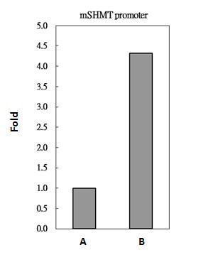
GTX103436 ChIP assay Image
c-Myc antibody immunoprecipitates c-Myc protein-DNA in ChIP experiments. ChIP Sample: 293T whole cell lysate/extract A. 5 ug preimmune rabbit IgG B. 5 ug of c-Myc antibody (GTX103436) The precipitated DNA was detected by PCR with primer set targeting to mSHMT promoter.
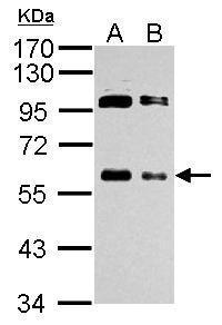
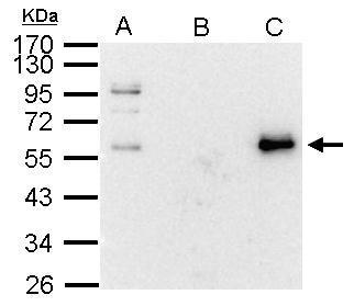
GTX103436 WB Image
Sample (whole cell lysate)
A: 293T 20ug
B: 293T 10ug
10% SDS PAGE
GTX103436 diluted at 1:1000
The HRP-conjugated anti-rabbit IgG antibody (GTX213110-01) was used to detect the primary antibody.
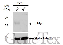
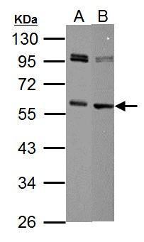
GTX103436 WB Image
Wild-type (WT) and c-Myc knockout (KO) 293T cell extracts (30 ug) were separated by 10% SDS-PAGE, and the membrane was blotted with c-Myc antibody (GTX103436) diluted at 1:2000. The HRP-conjugated anti-rabbit IgG antibody (GTX213110-01) was used to detect the primary antibody, and the signal was developed with Trident ECL plus-Enhanced.
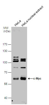
GTX103436 WB Image
c-Myc antibody detects c-Myc protein by western blot analysis. HeLa whole cell extracts and nuclear extracts (30 ug) were separated by 7.5% SDS-PAGE, and the membrane was blotted with c-Myc antibody (GTX103436) diluted at 1:1000. The HRP-conjugated anti-rabbit IgG antibody (GTX213110-01) was used to detect the primary antibody.