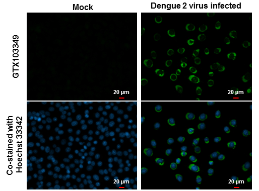
GTX103349 ICC/IF Image
NS4B (Dengue virus) antibody detects NS4B (Dengue virus) protein at cytoplasm by immunofluorescent analysis.
Samples: BHK-21 cells mock (left) and infected with Dengue virus 2 (right) were fixed in MeOH.
Green: NS4B (Dengue virus) protein stained by NS4B (Dengue virus) antibody (GTX103349) diluted at 1:2000.
Blue: Hoechst 33342 staining.
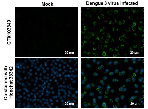
GTX103349 ICC/IF Image
NS4B (Dengue virus) antibody detects NS4B (Dengue virus) protein at cytoplasm by immunofluorescent analysis.
Samples: BHK-21 cells mock (left) and infected with Dengue virus 3 (right) were fixed in MeOH.
Green: NS4B (Dengue virus) protein stained by NS4B (Dengue virus) antibody (GTX103349) diluted at 1:2000.
Blue: Hoechst 33342 staining.
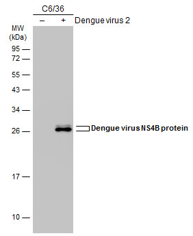
GTX103349 WB Image
Non-infected (?) and infected (+) C6/36 whole cell extracts (15 ug) were separated by 12% SDS-PAGE, and the membrane was blotted with Dengue virus NS4B protein antibody (GTX103349) diluted at 1:5000. The HRP-conjugated anti-rabbit IgG antibody (GTX213110-01) was used to detect the primary antibody.
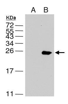
GTX103349 WB Image
Sample(15 ug of cell lysate)
A: BHK21
B: DV infected BHK21
10% SDS PAGE
GTX103349 diluted at 1:5000
The HRP-conjugated anti-rabbit IgG antibody (GTX213110-01) was used to detect the primary antibody.
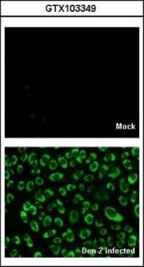
GTX103349 ICC/IF Image
GTX103349 IF Image Immunofluorescence analysis of Denque virus 2-infected BHK-21, using Non-structural protein 4B (Dengue virus 2 ) antibody (GTX103349) at 1:2000 dilution.
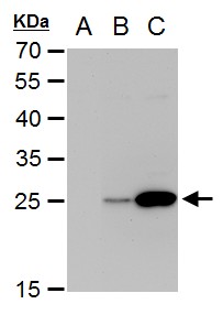
GTX103349 WB Image
NS4B (Dengue virus) antibody detects NS4B (Dengue virus) protein by western blot analysis.
A. 30 ug BHK-21 whole cell extrac
B. 30 ug whole cell extract of Dengue virus type 2 infected BHK-21 cells
C. 30 ug whole cell extract of Dengue virus type 3 infected BHK-21 cells
12% SDS-PAGE
NS4B (Dengue virus) antibody (GTX103349) dilution: 1:5000
The HRP-conjugated anti-rabbit IgG antibody (GTX213110-01) was used to detect the primary antibody.