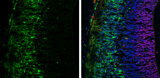
GTX102424 IHC-Fr Image
Tyrosine Hydroxylase antibody [N1C1] detects Tyrosine Hydroxylase protein expression by immunohistochemical analysis.
Sample: Frozen sectioned E13.5 Rat brain.
Green: Tyrosine Hydroxylase protein stained by Tyrosine Hydroxylase antibody [N1C1] (GTX102424) diluted at 1:250.
Red: SOX2, stained by SOX2 antibody [GT1876] (GTX627404) diluted at 1:250.
Blue: Fluoroshield with DAPI (GTX30920).
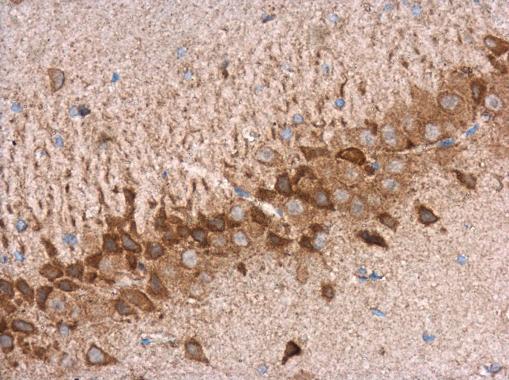
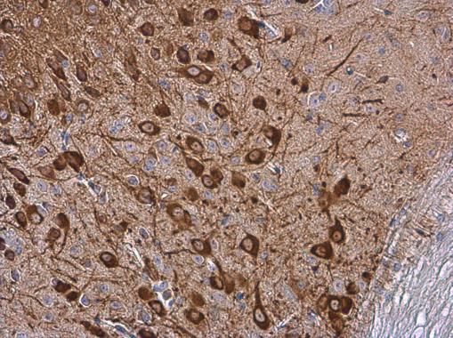
GTX102424 IHC-P Image
Tyrosine Hydroxylase antibody [N1C1] detects Tyrosine Hydroxylase protein at cytoplasm in rat brain by immunohistochemical analysis.
Sample: Paraffin-embedded rat brain.
Tyrosine Hydroxylase antibody [N1C1] (GTX102424) diluted at 1:500.
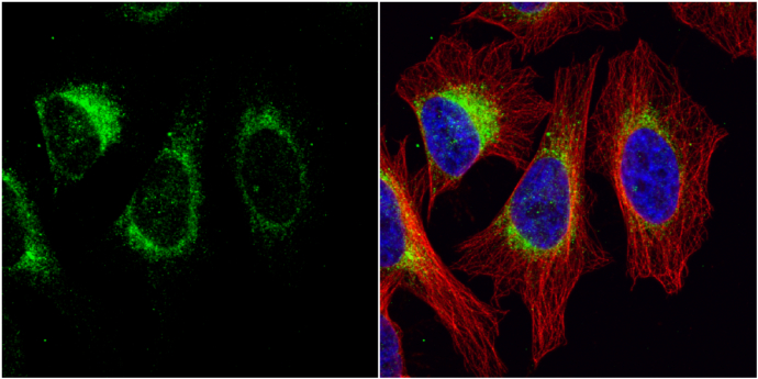
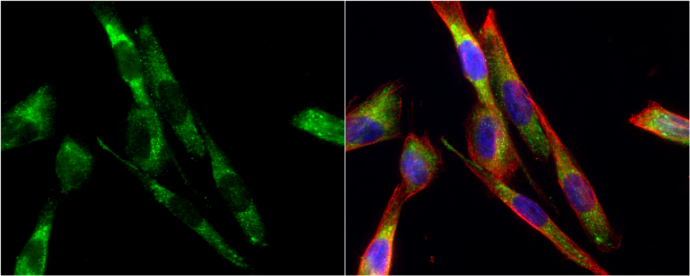
GTX102424 ICC/IF Image
Tyrosine Hydroxylase antibody [N1C1] detects Tyrosine Hydroxylase protein at cytoplasm by immunofluorescent analysis.
Sample: HeLa cells were fixed in 4% paraformaldehyde at RT for 15 min.
Green: Tyrosine Hydroxylase protein stained by Tyrosine Hydroxylase antibody [N1C1] (GTX102424) diluted at 1:200.
Red: alpha Tubulin, a cytoskeleton marker, stained by alpha Tubulin antibody [GT114] (GTX628802) diluted at 1:1000.
Blue: Hoechst 33342 staining.
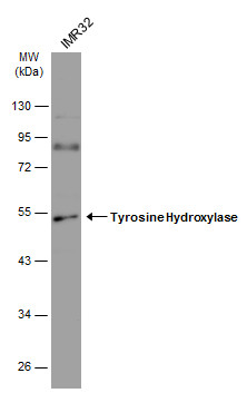
GTX102424 WB Image
Whole cell extract (30 ug) was separated by 10% SDS-PAGE, and the membrane was blotted with Tyrosine Hydroxylase antibody [N1C1] (GTX102424) diluted at 1:1000. The HRP-conjugated anti-rabbit IgG antibody (GTX213110-01) was used to detect the primary antibody.
GTX102424 ICC/IF Image
Tyrosine Hydroxylase antibody [N1C1] detects Tyrosine Hydroxylase protein at cytoplasm by immunofluorescent analysis.
Sample: SK-N-SH cells were fixed in 4% paraformaldehyde at RT for 15 min.
Green: Tyrosine Hydroxylase protein stained by Tyrosine Hydroxylase antibody [N1C1] (GTX102424) diluted at 1:200.
Red: alpha Tubulin, a cytoskeleton marker, stained by alpha Tubulin antibody [GT114] (GTX628802) diluted at 1:1000.
Blue: Hoechst 33342 staining.

GTX102424 IHC-P Image
Tyrosine Hydroxylase antibody [N1C1] detects Tyrosine Hydroxylase protein at cell membrane and cytoplasm by immunohistochemical analysis.Sample: Paraffin-embedded mouse brain.Tyrosine Hydroxylase stained by Tyrosine Hydroxylase antibody [N1C1] (GTX102424) diluted at 1:500.
GTX102424 IHC-P Image
Tyrosine Hydroxylase antibody [N1C1] detects Tyrosine Hydroxylase protein at cell membrane and cytoplasm by immunohistochemical analysis.Sample: Paraffin-embedded mouse brain.Tyrosine Hydroxylase stained by Tyrosine Hydroxylase antibody [N1C1] (GTX102424) diluted at 1:500.
GTX102424 IHC-P Image
Tyrosine Hydroxylase antibody [N1C1] detects Tyrosine Hydroxylase protein at cell membrane and cytoplasm by immunohistochemical analysis.Sample: Paraffin-embedded mouse brain.Tyrosine Hydroxylase stained by Tyrosine Hydroxylase antibody [N1C1] (GTX102424) diluted at 1:500.
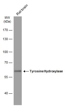
GTX102424 WB Image
Rat tissue extract (50 ug) was separated by 7.5% SDS-PAGE, and the membrane was blotted with Tyrosine Hydroxylase antibody (GTX102424) diluted at 1:1000. The HRP-conjugated anti-rabbit IgG antibody (GTX213110-01) was used to detect the primary antibody.
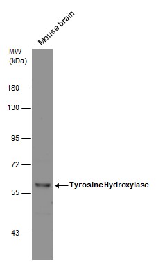
GTX102424 WB Image
Mouse tissue extract (50 ug) was separated by 7.5% SDS-PAGE, and the membrane was blotted with Tyrosine Hydroxylase antibody [N1C1] (GTX102424) diluted at 1:1000.
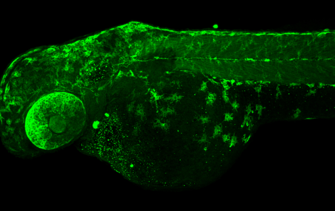
GTX102424 IHC-Wm Image
Tyrosine Hydroxylase antibody [N1C1] detects Tyrosine Hydroxylase protein on zebrafish by whole mount immunohistochemical analysis.
Sample: 2 days-post-fertilization zebrafish embryo.
Tyrosine Hydroxylase antibody [N1C1] (GTX102424) dilution: 1:100.
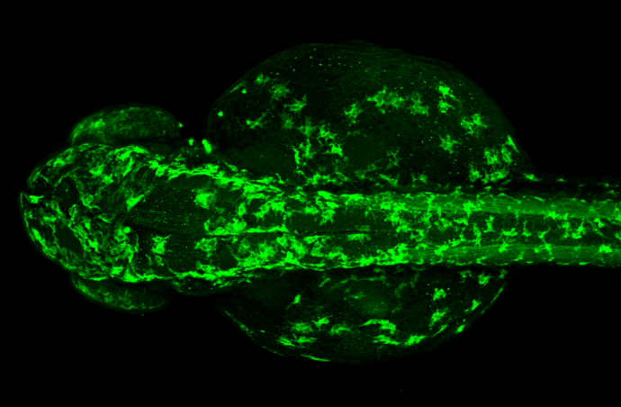
GTX102424 IHC-Wm Image
Tyrosine Hydroxylase antibody [N1C1] detects Tyrosine Hydroxylase protein on zebrafish by whole mount immunohistochemical analysis.
Sample: 2 days-post-fertilization zebrafish embryo.
Tyrosine Hydroxylase antibody [N1C1] (GTX102424) dilution: 1:100.
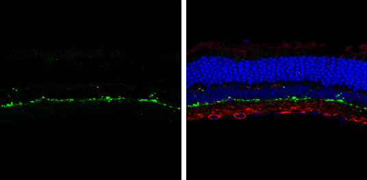
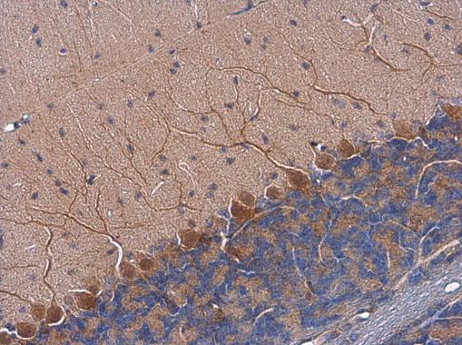
GTX102424 IHC-Fr Image
Tyrosine Hydroxylase antibody [N1C1] detects Tyrosine Hydroxylase protein by immunohistochemical analysis.
Sample: Frozen sectioned adult mouse retina.
Green: Tyrosine Hydroxylase protein stained by Tyrosine Hydroxylase antibody [N1C1] (GTX102424) diluted at 1:250.
Red: beta Tubulin 3/ TUJ1, stained by beta Tubulin 3/ TUJ1 antibody [GT11710] (GTX631836) diluted at 1:250.
Blue: Fluoroshield with DAPI (GTX30920).