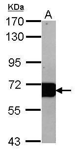
GTX102419 WB Image
Sample (20 ug of whole cell lysate)
A: mouse Liver
7.5% SDS PAGE
GTX102419 diluted at 1:10000
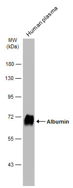
GTX102419 WB Image
Human plasma (1 ug) was separated by 7.5% SDS-PAGE, and the membrane was blotted with Albumin antibody (GTX102419) diluted at 1:100000. The HRP-conjugated anti-rabbit IgG antibody (GTX213110-01) was used to detect the primary antibody.
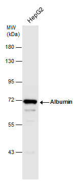
GTX102419 WB Image
Whole cell extract (30 ug) was separated by 7.5% SDS-PAGE, and the membrane was blotted with Albumin antibody (GTX102419) diluted at 1:1000.
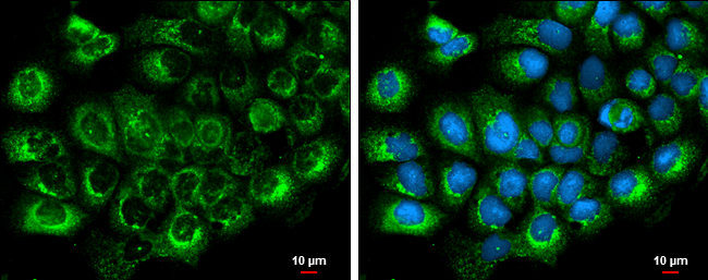
GTX102419 ICC/IF Image
Albumin antibody detects Albumin protein at cytoplasm by immunofluorescent analysis.
Sample: A431 cells were fixed in ice-cold MeOH for 5 min.
Green: Albumin protein stained by Albumin antibody (GTX102419) diluted at 1:500.
Blue: Hoechst 33342 staining.
Scale bar = 10 um.
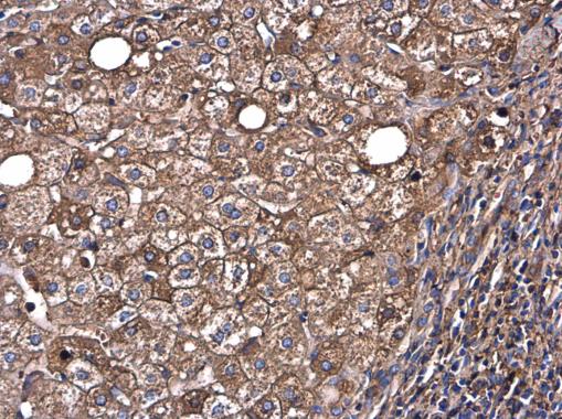
GTX102419 IHC-P Image
Albumin antibody detects Albumin protein at cytoplasm in human hepatocellular carcinoma by immunohistochemical analysis.
Sample: Paraffin-embedded human hepatocellular carcinoma.
Albumin antibody (GTX102419) diluted at 1:500.
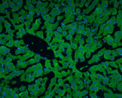
GTX102419 IHC-Fr Image
Albumin antibody detects Albumin protein at cytoplasm in rat liver by immunohistochemical analysis.
Sample: Frozen section of rat liver.
Albumin antibody (GTX102419) diluted at 1:200.