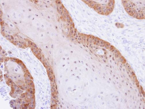
GTX102229 IHC-P Image
Immunohistochemical analysis of paraffin-embedded Cal27 xenograft, using XBP1(GTX102229) antibody at 1:500 dilution.
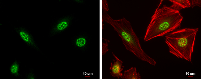
GTX102229 ICC/IF Image
XBP1 antibody [N3C3] detects XBP1 protein at nucleus by immunofluorescent analysis.
Sample: HeLa cells were fixed in 4% paraformaldehyde at RT for 15 min.
Green: XBP1 protein stained by XBP1 antibody [N3C3] (GTX102229) diluted at 1:500.
Red: phalloidin, a cytoskeleton marker, stained by () diluted at 1:200.
Blue: Hoechst 33342 staining.
Scale bar = 10 um.
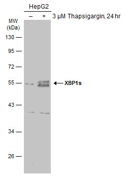
GTX102229 WB Image
Untreated (?) and treated (+) HepG2 whole cell extracts (30 ug) were separated by 10% SDS-PAGE, and the membrane was blotted with XBP1 antibody [N3C3] (GTX102229) diluted at 1:1000. The HRP-conjugated anti-rabbit IgG antibody (GTX213110-01) was used to detect the primary antibody.
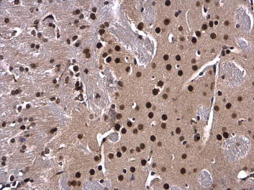
GTX102229 IHC-P Image
XBP1 antibody [N3C3] detects XBP1 protein at nucleus in mouse brain by immunohistochemical analysis.
Sample: Paraffin-embedded mouse brain.
XBP1 antibody [N3C3] (GTX102229) diluted at 1:500.
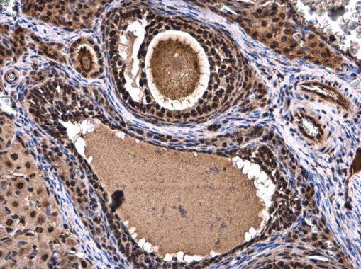
GTX102229 IHC-P Image
XBP1 antibody [N3C3] detects XBP1 protein at nucleus in rat ovary by immunohistochemical analysis.
Sample: Paraffin-embedded rat ovary.
XBP1 antibody [N3C3] (GTX102229) diluted at 1:500.
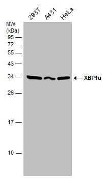
GTX102229 WB Image
Various whole cell extracts (30 ug) were separated by 12% SDS-PAGE, and the membrane was blotted with XBP1 antibody [N3C3] (GTX102229) diluted at 1:1000.