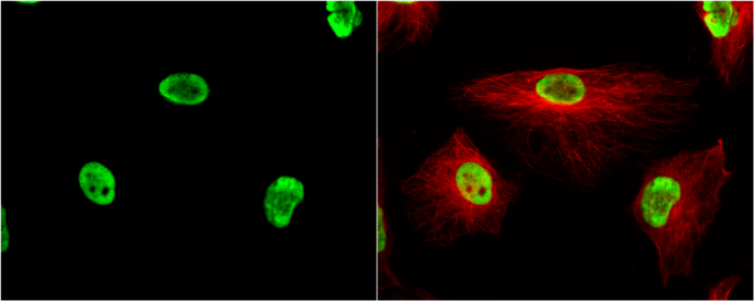
GTX102226 ICC/IF Image
KAP1 antibody [N3C2], Internal detects KAP1 protein at nucleus by immunofluorescent analysis.
Sample: HeLa cells were fixed in 4% paraformaldehyde at RT for 15 min.
Green: KAP1 protein stained by KAP1 antibody [N3C2], Internal (GTX102226) diluted at 1:200.
Red: alpha Tubulin, a cytoskeleton marker, stained by alpha Tubulin antibody [B-5-1-2] (GTX11304) diluted at 1:10000.
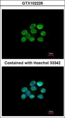
GTX102226 ICC/IF Image
Immunofluorescence analysis of paraformaldehyde-fixed A431, using KAP1(GTX102226) antibody at 1:200 dilution.
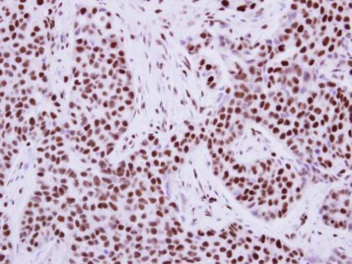
GTX102226 IHC-P Image
Immunohistochemical analysis of paraffin-embedded human breast cancer, using KAP1(GTX102226) antibody at 1:500 dilution.
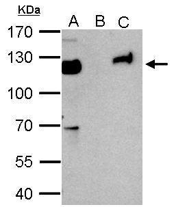
GTX102226 IP Image
KAP1 antibody immunoprecipitates KAP1 protein in IP experiments. IP Sample: 1000 ug HeLa whole cell lysate/extract A. 50 ug HeLa whole cell lysate/extract B. Control with 2 ug of preimmune rabbit IgG C. Immunoprecipitation of KAP1 protein by 2 ug of KAP1 antibody (GTX102226) 7.5% SDS-PAGE The immunoprecipitated KAP1 protein was detected by KAP1 antibody (GTX102226) diluted at 1:1000. EasyBlot anti-rabbit IgG (GTX221666-01) was used as a secondary reagent.
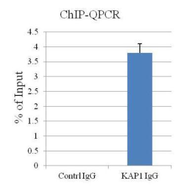
GTX102226 ChIP assay Image
ChIP assay followed by QPCR on a known KAP1-binding region
within the ZNF180 3ÅfUTR (Mol Cell Biol, 2011, Lyengar et al)
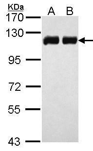
GTX102226 WB Image
Sample (30 ug of whole cell lysate)
A: H1299
B: HeLa
7.5% SDS PAGE
GTX102226 diluted at 1:10000
The HRP-conjugated anti-rabbit IgG antibody (GTX213110-01) was used to detect the primary antibody.
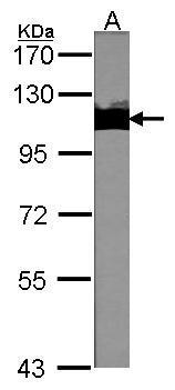
GTX102226 WB Image
Sample (30 ug of whole cell lysate)
A:NIH-3T3
7.5% SDS PAGE
GTX102226 diluted at 1:1000
The HRP-conjugated anti-rabbit IgG antibody (GTX213110-01) was used to detect the primary antibody.