GTX102150 ICC/IF Image
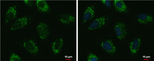
UQCRFS1 antibody [N1C3] detects UQCRFS1 protein at mitochondria by immunofluorescent analysis.
Sample: A549 cells were fixed in 2% paraformaldehyde/culture medium at 37Åé for 30 min.
Green: UQCRFS1 protein stained by UQCRFS1 antibody [N1C3] (GTX102150) diluted at 1:500.
Blue: Hoechst 33342 staining.
Scale bar = 10 um.
GTX102150 ICC/IF Image
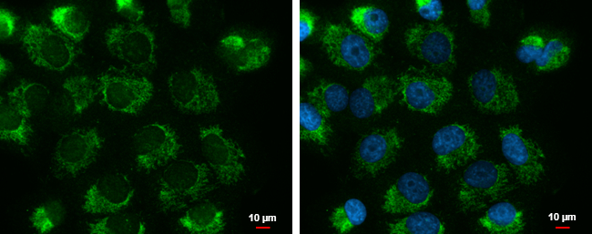
UQCRFS1 antibody [N1C3] detects UQCRFS1 protein at mitochondria by immunofluorescent analysis.
Sample: H1299 cells were fixed in 2% paraformaldehyde/culture medium at 37oC for 30 min.
Green: UQCRFS1 protein stained by UQCRFS1 antibody [N1C3] (GTX102150) diluted at 1:500.
Blue: Hoechst 33342 staining.
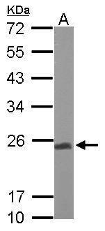
GTX102150 WB Image
Sample (50 ug of whole cell lysate)
A: Mouse brain
12% SDS PAGE
GTX102150 diluted at 1:1000
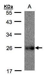
GTX102150 WB Image
Sample(30 ug whole cell lysate)
A: Raji(GTX27908)
12% SDS PAGE
GTX102150 diluted at 1:500