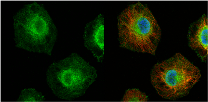
GTX102026 ICC/IF Image
MCL1 antibody detects MCL1 protein at cytoplasm and nucleus by immunofluorescent analysis.
Sample: HeLa cells were fixed in 4% paraformaldehyde at RT for 15 min.
Green: MCL1 protein stained by MCL1 antibody (GTX102026) diluted at 1:200.
Red: alpha Tubulin, a cytoskeleton marker, stained by alpha Tubulin antibody [B-5-1-2] (GTX11304) diluted at 1:10000.
Blue: Hoechst 33342 staining.
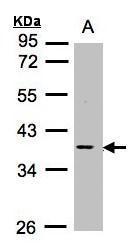
GTX102026 WB Image
Sample (30ug whole cell lysate)
A:Raji (GTX27908)
10% SDS PAGE
GTX102026 diluted at 1:1000
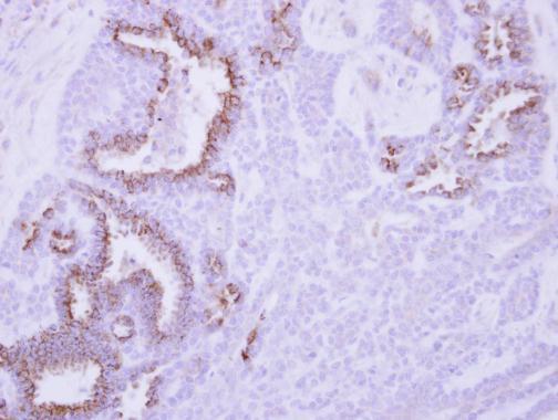
GTX102026 IHC-P Image
Immunohistochemical analysis of paraffin-embedded human breast cancer, using MCL1(GTX102026) antibody at 1:250 dilution.
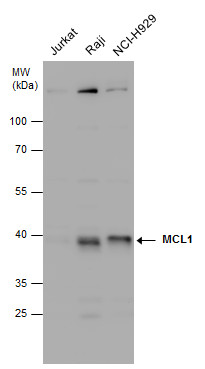
GTX102026 WB Image
MCL1 antibody detects MCL1 protein by Western blot analysis. Various whole cell extracts (30 ug) were separated by 10% SDS-PAGE, and the membrane was blotted with MCL1 antibody (GTX102026) diluted at 1:1000.
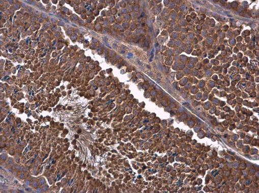
GTX102026 IHC-P Image
MCL1 antibody detects MCL1 protein at cytoplasm in mouse testis by immunohistochemical analysis.
Sample: Paraffin-embedded mouse testis.
MCL1 antibody (GTX102026) diluted at 1:500.