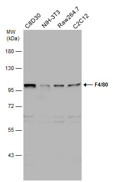
GTX101895 WB Image
Various whole cell extracts (30 ug) were separated by 7.5% SDS-PAGE, and the membrane was blotted with F4/80 antibody [C2C3], C-term (GTX101895) diluted at 1:1000. The HRP-conjugated anti-rabbit IgG antibody (GTX213110-01) was used to detect the primary antibody.
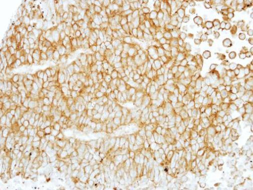
GTX101895 IHC-P Image
Immunohistochemical analysis of paraffin-embedded PC13 xenograft, using EMR1(GTX101895) antibody at 1:100 dilution.

GTX101895 WB Image
EMR1 antibody [C2C3], C-term detects EMR1 protein by western blot analysis.
A. 30 ug Jurkat whole cell lysate/extract
B. 30 ug Raji whole cell lysate/extract
C. 30 ug K562 whole cell lysate/extract
7.5% SDS-PAGE
EMR1 antibody [C2C3], C-term (GTX101895) dilution: 1:1000
The HRP-conjugated anti-rabbit IgG antibody (GTX213110-01) was used to detect the primary antibody.
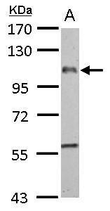
GTX101895 WB Image
EMR1 antibody [C2C3], C-term detects EMR1 protein by western blot analysis.
A. 50 ug rat liver lysate/extract
7.5% SDS-PAGE
EMR1 antibody [C2C3], C-term (GTX101895) dilution: 1:1000
The HRP-conjugated anti-rabbit IgG antibody (GTX213110-01) was used to detect the primary antibody.