GTX101810 IHC-P Image
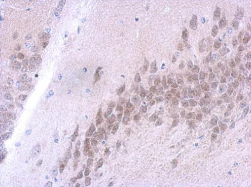
FUS antibody [C1C3] detects FUS protein at nucleus on mouse brain by immunohistochemical analysis.
Sample: Paraffin-embedded mouse brain.
FUS antibody [C1C3] (GTX101810) dilution: 1:500.
GTX101810 IHC-P Image
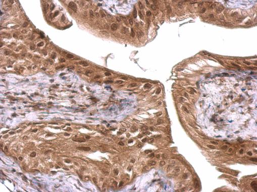
FUS antibody [C1C3] detects FUS protein at nucleus on mouse urinary bladder by immunohistochemical analysis.
Sample: Paraffin-embedded mouse urinary bladder.
FUS antibody [C1C3] (GTX101810) dilution: 1:500.
GTX101810 WB Image
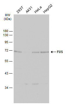
Various whole cell extracts (30 ug) were separated by 10% SDS-PAGE, and the membrane was blotted with FUS antibody [C1C3] (GTX101810) diluted at 1:1000. The HRP-conjugated anti-rabbit IgG antibody (GTX213110-01) was used to detect the primary antibody.
GTX101810 WB Image
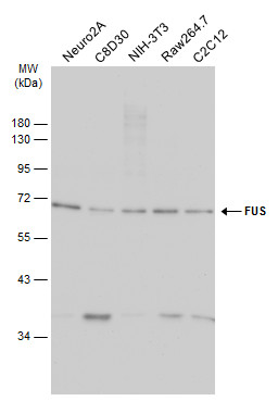
Various whole cell extracts (30 ug) were separated by 10% SDS-PAGE, and the membrane was blotted with FUS antibody [C1C3] (GTX101810) diluted at 1:1000. The HRP-conjugated anti-rabbit IgG antibody (GTX213110-01) was used to detect the primary antibody.