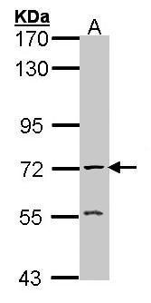
GTX101648 WB Image
Sample (30 ug of whole cell lysate)
A: Raji
7.5% SDS PAGE
GTX101648 diluted at 1:1000
The HRP-conjugated anti-rabbit IgG antibody (GTX213110-01) was used to detect the primary antibody.
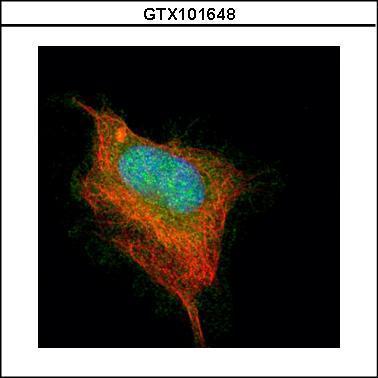
GTX101648 ICC/IF Image
Confocal immunofluorescence analysis (Olympus FV10i) of paraformaldehyde-fixed HeLa, using AChE(GTX101648) antibody (Green) at 1:500 dilution. Alpha-tubulin filaments were labeled with GTX11304 (Red) at 1:2500.
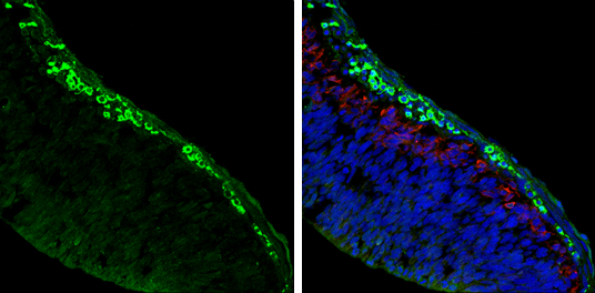
GTX101648 IHC-Fr Image
AChE antibody detects AChE protein expression by immunohistochemical analysis.
Sample: Frozen sectioned E13.5 Rat brain.
Green: AChE protein stained by AChE antibody (GTX101648) diluted at 1:250.
Red: beta Tubulin 3/ TUJ1, a mature neuron marker, stained by beta Tubulin 3/ TUJ1 antibody [GT11710] (GTX631836) diluted at 1:500.
Blue: Fluoroshield with DAPI (GTX30920).
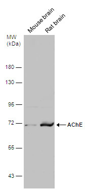
GTX101648 WB Image
Various tissue extracts (50 ug) were separated by 7.5% SDS-PAGE, and the membrane was blotted with AChE antibody (GTX101648) diluted at 1:500. The HRP-conjugated anti-rabbit IgG antibody (GTX213110-01) was used to detect the primary antibody.
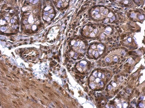
GTX101648 IHC-P Image
AChE antibody detects AChE protein at nucleus on mouse colon by immunohistochemical analysis.
Sample: Paraffin-embedded mouse colon.
AChE antibody (GTX101648) dilution: 1:500.
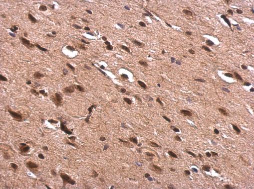
GTX101648 IHC-P Image
AChE antibody detects AChE protein at nucleus on rat fore brain by immunohistochemical analysis.
Sample: Paraffin-embedded rat fore brain.
AChE antibody (GTX101648) dilution: 1:500.
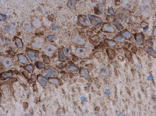
GTX101648 IHC-P Image
AChE antibody detects AChE protein at cytoplasm and nucleus in rat brain by immunohistochemical analysis.
Sample: Paraffin-embedded rat brain.
AChE antibody (GTX101648) diluted at 1:500.
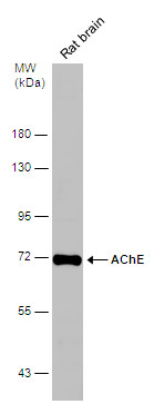
GTX101648 WB Image
Rat tissue extract (50 ug) was separated by 7.5% SDS-PAGE, and the membrane was blotted with AChE antibody (GTX101648) diluted at 1:500.