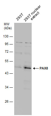
GTX101583 WB Image
293T whole cell and nuclear extracts (30 ug) were separated by 10% SDS-PAGE, and the membrane was blotted with PAX8 antibody (GTX101583) diluted at 1:1000.
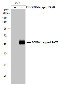
GTX101583 WB Image
Non-transfected (?) and transfected (+) 293T whole cell extracts (30 ug) were separated by 10% SDS-PAGE, and the membrane was blotted with PAX8 antibody (GTX101583) diluted at 1:5000. The HRP-conjugated anti-rabbit IgG antibody (GTX213110-01) was used to detect the primary antibody.
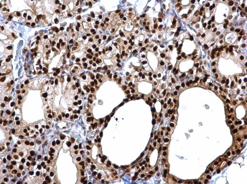
GTX101583 IHC-P Image
PAX8 antibody detects PAX8 protein at cytosol and nucleus on rat thyroid gland by immunohistochemical analysis.
Sample: Paraffin-embedded rat thyroid gland.
PAX8 antibody (GTX101583) dilution: 1:500.
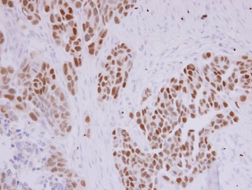
GTX101583 IHC-P Image
PAX8 antibody detects PAX8 protein at nucleolus by immunohistochemical analysis.
Sample: Paraffin-embedded DLD1 xenograft .
PAX8 antibody (GTX101583) diluted at 1:500.
GTX101583 IHC-P Image
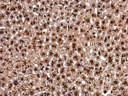
PAX8 antibody detects PAX8 protein at nucleus in mouse liver by immunohistochemical analysis.
Sample: Paraffin-embedded mouse liver.
PAX8 antibody (GTX101583) diluted at 1:500.
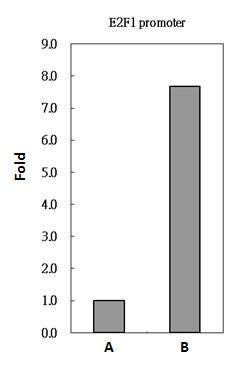
GTX101583 ChIP assay Image
PAX8 antibody immunoprecipitates PAX8 protein-DNA in ChIP experiments. ChIP Sample: 293T whole cell lysate/extract A. 5 ug preimmune rabbit IgG B. 5 ug of PAX8 antibody (GTX101583) The precipitated DNA was detected by PCR with primer set targeting to E2F1 promoter.
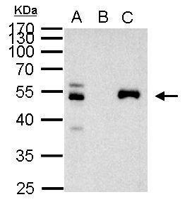
GTX101583 IP Image
PAX8 antibody immunoprecipitates PAX8 protein in IP experiments. IP Sample: 1000 ug 293T whole cell lysate/extract A. 40 ug 293T whole cell lysate/extract B. Control with 2 ug of preimmune rabbit IgG C. Immunoprecipitation of PAX8 protein by 2 ug of PAX8 antibody (GTX101583) 10% SDS-PAGE The immunoprecipitated PAX8 protein was detected by PAX8 antibody (GTX101583) diluted at 1:1000. EasyBlot anti-rabbit IgG (GTX221666-01) was used as a secondary reagent.
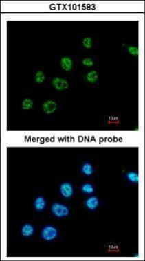
GTX101583 ICC/IF Image
Immunofluorescence analysis of paraformaldehyde-fixed A549, using PAX8(GTX101583) antibody at 1:200 dilution.