GTX101553 IHC-Fr Image
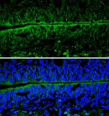
NSE antibody [N1C1] detects NSE protein expression by immunohistochemical analysis.
Sample: Frozen sectioned E13.5 Rat brain.
Green: NSE protein stained by NSE antibody [N1C1] (GTX101553) diluted at 1:250.
Blue: Fluoroshield with DAPI (GTX30920).
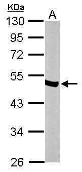
GTX101553 WB Image
Sample (50 ug of whole cell lysate)
A: mouse brain
10% SDS PAGE
GTX101553 diluted at 1:10000
The HRP-conjugated anti-rabbit IgG antibody (GTX213110-01) was used to detect the primary antibody.
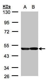
GTX101553 WB Image
Sample(30 ug of whole cell lysate)
A:A431(GTX27909)
B:H1299
10% SDS PAGE
GTX101553 diluted at 1:2000
The HRP-conjugated anti-rabbit IgG antibody (GTX213110-01) was used to detect the primary antibody.
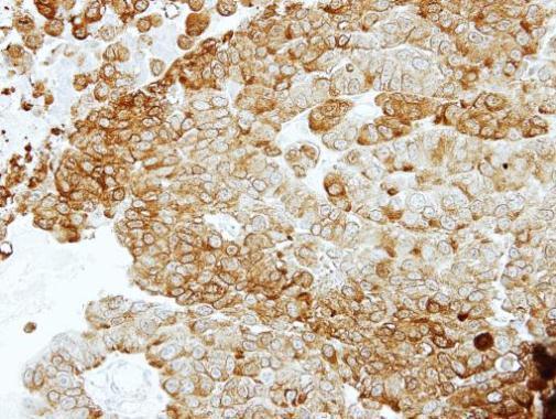
GTX101553 IHC-P Image
Immunohistochemical analysis of paraffin-embedded PC14 xenograft, using ENO2(GTX101553) antibody at 1:100 dilution.