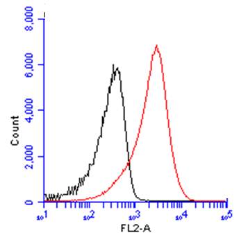
GTX101512 FACS Image
CD19 antibody [C1C3] (GTX101512) detects CD19 protein by flow cytometry analysis.
Sample: mouse splenocytes cell fixed in 4% paraformaldehyde at 4oC for 15 min.
Black: Unlabelled sample was used as a control.
Red: CD19 antibody [C1C3] (GTX101512) dilution: 1:50.
Acquisition of 20,000 events were collected using a Dylight 488-conjugated secondary antibody for FACS analysis.
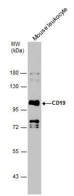
GTX101512 WB Image
Mouse tissue extract (50 ug) was separated by 7.5% SDS-PAGE, and the membrane was blotted with CD19 antibody [C1C3] (GTX101512) diluted at 1:1000.
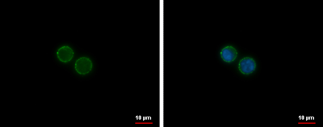
GTX101512 ICC/IF Image
CD19 antibody [C1C3] detects CD19 protein at membrane by immunofluorescent analysis.
Sample: Raji cells were fixed in ice-cold MeOH for 5 min.
Green: CD19 protein stained by CD19 antibody [C1C3] (GTX101512) diluted at 1:1000.
Blue: Hoechst 33342 staining.
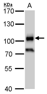
GTX101512 WB Image
CD19 antibody detects CD19 protein by western blot analysis.
A. 30 ug Raji whole cell lysate/extract
7.5 % SDS-PAGE
CD19 antibody (GTX101512) dilution: 1:1000