GTX101507 IHC-P Image
SOX2 antibody [N1C3] detects SOX2 protein at nucleus on mouse fore brain by immunohistochemical analysis.
Sample: Paraffin-embedded mouse fore brain.
SOX2 antibody [N1C3] (GTX101507) diluted at 1:500.
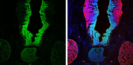
GTX101507 IHC-Fr Image
SOX2 antibody [N1C3] detects SOX2 protein at nucleus by immunohistochemical analysis.
Sample: Frozen sectioned E13.5 rat brain.
Green: SOX2 protein stained by SOX2 antibody [N1C3] (GTX101507) diluted at 1:250.
Red: beta Tubulin 3/ TUJ1, a mature neuron marker, stained by beta Tubulin 3/ TUJ1 antibody [GT11710] (GTX631836) diluted at 1:250.
Blue: Fluoroshield with DAPI (GTX30920).
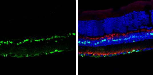
GTX101507 IHC-Fr Image
SOX2 antibody [N1C3] detects SOX2 protein at nucleus by immunohistochemical analysis.
Sample: Frozen sectioned adult mouse retina.
Green: SOX2 protein stained by SOX2 antibody [N1C3] (GTX101507) diluted at 1:250.
Red: Protein kinase C alpha staining.
Blue: Fluoroshield with DAPI (GTX30920).
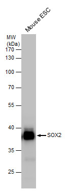
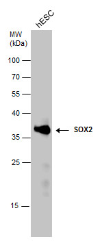
GTX101507 WB Image
SOX2 antibody detects SOX2 protein by western blot analysis. Mouse ESC lysate (50 ug) was separated by 10% SDS-PAGE, and the membrane was blotted with SOX2 antibody (GTX101507) at a dilution of 1:2500. The HRP-conjugated anti-rabbit IgG antibody (GTX213110-01) was used to detect the primary antibody.
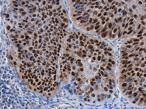
GTX101507 IHC-P Image
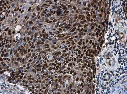
SOX2 antibody [N1C3] detects SOX2 protein at nucleus in human cervical carcinoma by immunohistochemical analysis.
Sample: Paraffin-embedded human cervical carcinoma.
SOX2 antibody [N1C3] (GTX101507) diluted at 1:500.
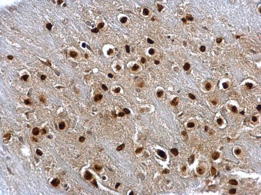
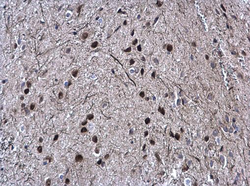
GTX101507 IHC-P Image
SOX2 antibody [N1C3] detects SOX2 protein at nucleus on rat brain stem by immunohistochemical analysis.
Sample: Paraffin-embedded rat brain stem.
SOX2 antibody [N1C3] (GTX101507) dilution: 1:500.
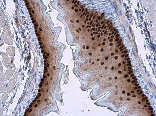
GTX101507 IHC-P Image
SOX2 antibody [N1C3] detects SOX2 protein at nucleus in mouse esophagus by immunohistochemical analysis.
Sample: Paraffin-embedded mouse esophagus.
SOX2 antibody [N1C3] (GTX101507) diluted at 1:500.
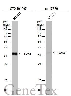
GTX101507 WB Image
Whole cell extract (30 ug) was separated by 12% SDS-PAGE, and the membranes were blotted with SOX2 antibody [N1C3] (GTX101507) diluted at 1:1000 and competitor's antibody (sc-17320) diluted at 1:500. The HRP-conjugated anti-rabbit IgG antibody (GTX213110-01) was used to detect the primary antibody.
*The competitor is not affiliated with GeneTex and does not endorse this product.
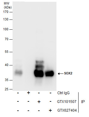
GTX101507 IP Image
Immunoprecipitation of SOX2 protein from NT2D1 whole cell extracts using 5 ug of SOX2 antibody [N1C3] (GTX101507) or SOX2 antibody [GT1876] (GTX627404).
Western blot analysis was performed using SOX2 antibody [N1C3] (GTX101507).
EasyBlot anti-Rabbit IgG (GTX221666-01) was used as a secondary reagent.
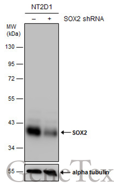
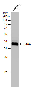
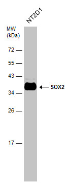
GTX101507 WB Image
Non-transfected (?) and transfected (+) NT2D1 whole cell extracts (30 ug) were separated by 10% SDS-PAGE, and the membrane was blotted with SOX2 antibody [N1C3] (GTX101507) diluted at 1:15000. The HRP-conjugated anti-rabbit IgG antibody (GTX213110-01) was used to detect the primary antibody.
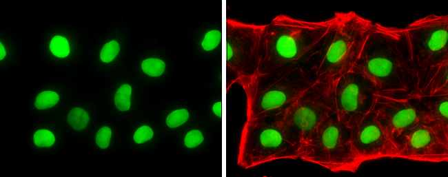
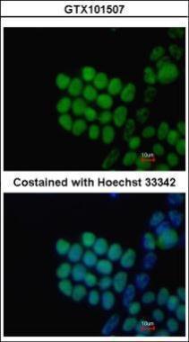
GTX101507 ICC/IF Image
SOX2 antibody [N1C3] detects SOX2 protein at nucleus by immunofluorescent analysis.Sample: NT2D1 cells were fixed in 4% paraformaldehyde at RT for 15 min.Green: SOX2 stained by SOX2 antibody [N1C3] (GTX101507) diluted at 1:500.Red: phalloidin, a cytoskeleton marker, diluted at 1:100.
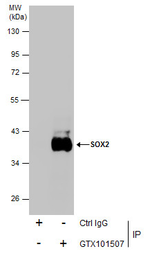
GTX101507 IP Image
Immunoprecipitation of SOX2 protein from NT2D1 whole cell extracts using 5 ug of SOX2 antibody [N1C3] (GTX101507).
Western blot analysis was performed using SOX2 antibody [N1C3] (GTX101507).
EasyBlot anti-Rabbit IgG (GTX221666-01) was used as a secondary reagent.
GTX101507 WB Image
SOX2 antibody detects SOX2 protein by Western blot analysis. Whole cell extracts (30 ug) was separated by 12% SDS-PAGE, and the membrane was blotted with SOX2 antibody (GTX101507) at a dilution of 1:2500.
GTX101507 IHC-P Image
SOX2 antibody [N1C3] detects SOX2 protein at nucleus in mouse fetal brain by immunohistochemical analysis.
Sample: Paraffin-embedded mouse fetal brain.
Green: SOX2 antibody [N1C3] (GTX101507) diluted at 1:200. The signal was developed using goat anti-rabbit IgG antibody (Dylight488) (GTX213110-04).
Blue: Nuclear staining with Hoechst 33342.
GTX101507 IHC-P Image
SOX2 antibody [N1C3] detects SOX2 protein at nucleus in human esophageal carcinoma by immunohistochemical analysis.
Sample: Paraffin-embedded human esophageal carcinoma.
SOX2 antibody [N1C3] (GTX101507) diluted at 1:500.
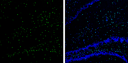
GTX101507 IHC-Fr Image
SOX2 antibody [N1C3] detects SOX2 protein expression by immunohistochemical analysis.
Sample: Frozen-sectioned adult mouse hippocampus.
Green: SOX2 protein stained by SOX2 antibody [N1C3] (GTX101507) diluted at 1:250.
Blue: Fluoroshield with DAPI (GTX30920).
GTX101507 WB Image
Whole cell extract (30 ug) was separated by 12% SDS-PAGE, and the membrane was blotted with SOX2 antibody [N1C3] (GTX101507) diluted at 1:10000.
GTX101507 WB Image
Whole cell extract (30 ug) was separated by 12% SDS-PAGE, and the membrane was blotted with SOX2 antibody [GT1876] (GTX101507) diluted at 1:10000.
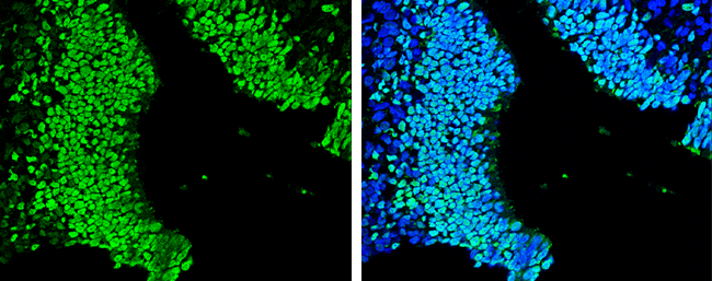
GTX101507 ICC/IF Image
Immunofluorescence analysis of paraformaldehyde-fixed mouse ESC, using SOX2(GTX101507) antibody at 1:80 dilution.
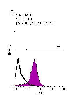
GTX101507 FACS Image
Flow cytometry on human embryonic stem cells, staining with SOX2 (GTX101507)antibody at 1:100 dilution(purple) or rabbit IgG (black).