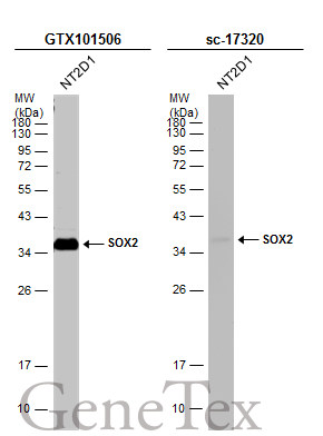
GTX101506 WB Image
Whole cell extract (30 ug) was separated by 12% SDS-PAGE, and the membranes were blotted with SOX2 antibody (GTX101506) diluted at 1:1000 and competitor's antibody (sc-17320) diluted at 1:500. The HRP-conjugated anti-rabbit IgG antibody (GTX213110-01) was used to detect the primary antibody, and the signal was developed with Trident ECL plus-Enhanced.
*The competitor is not affiliated with GeneTex and does not endorse this product.
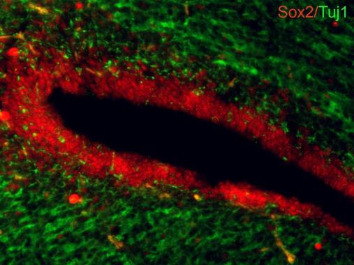
GTX101506 IHC-Fr Image
Sox2 antibodies detects Sox2 proteins on embryonic mouse brain by immunohistochemical analysis.
Sample:Frozen section of embryonic mouse brain (mE18.5).
Green: beta III Tubulin antibody [GT886] (GTX631830) diluted at 1:2000.
Red: Sox2 antibody (GTX101506) diluted at 1:250.
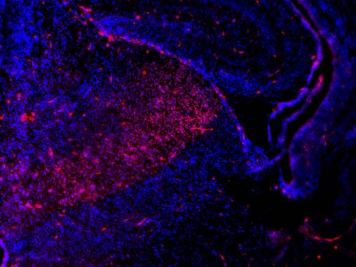
GTX101506 IHC-Fr Image
Sox2 antibodies detects Sox2 proteins on embryonic mouse brain by immunohistochemical analysis.
Sample:Frozen section of embryonic mouse brain (mE18.5). Red: Sox2 antibody (GTX101506) diluted at 1:500.
Blue: DAPI
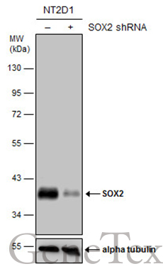
GTX101506 WB Image
Non-transfected (?) and transfected (+) NT2D1 whole cell extracts (30 ug) were separated by 10% SDS-PAGE, and the membrane was blotted with SOX2 antibody (GTX101506) diluted at 1:1000. The HRP-conjugated anti-rabbit IgG antibody (GTX213110-01) was used to detect the primary antibody.
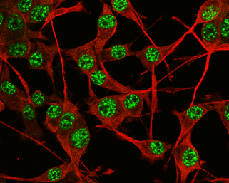
GTX101506 ICC/IF Image
SOX2 antibody detects SOX2 protein at nucleus by immunofluorescent analysis.
Sample: U-87 MG cells were fixed in 4% paraformaldehyde at RT for 15 min.
Green: SOX2 protein stained by SOX2 antibody (GTX101506) diluted at 1:200.
Red: beta Tubulin 3/ TUJ1 protein stained by beta Tubulin 3/ TUJ1 antibody (GTX631836) diluted at 1:200.
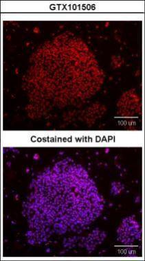
GTX101506 ICC/IF Image
Immunofluorescence analysis of paraformaldehyde-fixed mouse ESC, using SOX2(GTX101506) antibody at 1:200 dilution.
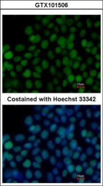
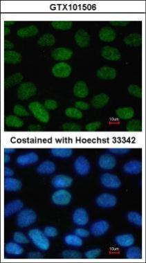
GTX101506 ICC/IF Image
Immunofluorescence analysis of paraformaldehyde-fixed human embryonic stem cell, using SOX2(GTX101506) antibody at 1:100 dilution.
GTX101506 WB Image
Sample (20 ug of whole cell lysate)
A: Human ESC
10% SDS PAGE
GTX101506 diluted at 1:1000
The HRP-conjugated anti-rabbit IgG antibody (GTX213110-01) was used to detect the primary antibody.
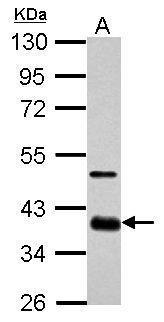
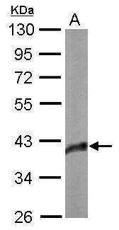
GTX101506 WB Image
Sample (30 ug of whole cell lysate)
A: GL261
10% SDS PAGE
GTX101506 diluted at 1:1000
The HRP-conjugated anti-rabbit IgG antibody (GTX213110-01) was used to detect the primary antibody.
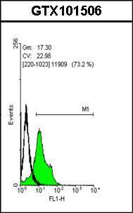
GTX101506 FACS Image
Flow cytometry on human embryonic stem cells, staining with SOX2 (GTX101506)antibody at 1:50 dilution(green) or rabbit IgG (black).
GTX101506 ICC/IF Image
Immunofluorescence analysis of paraformaldehyde-fixed Human ESC, using SOX2(GTX101506) antibody at 1:100 dilution.
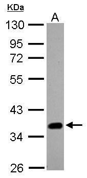
GTX101506 WB Image
Sample (20 ug of whole cell lysate )
A: A431 (GTX27909)
10% SDS PAGE
GTX101506 diluted at 1:1000
The HRP-conjugated anti-rabbit IgG antibody (GTX213110-01) was used to detect the primary antibody.