GTX101495 IHC-P Image
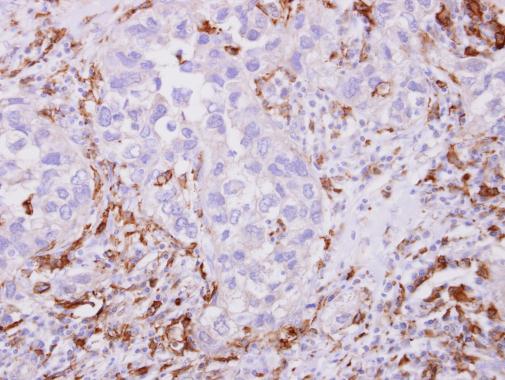
Iba1 antibody detects Iba1 protein at cytoplasm on human breast cancer stroma by immunohistochemical analysis.
Sample: Paraffin-embedded breast cancer stroma.
Iba1 antibody (GTX101495) dilution: 1:500.
GTX101495 IP Image
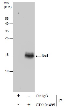
Immunoprecipitation of Iba1 protein from K562 whole cell extracts using 5 ug of Iba1 antibody (GTX101495).
Western blot analysis was performed using Iba1 antibody (GTX101495).
EasyBlot anti-Rabbit IgG (GTX221666-01) was used as a secondary reagent.
GTX101495 FACS Image
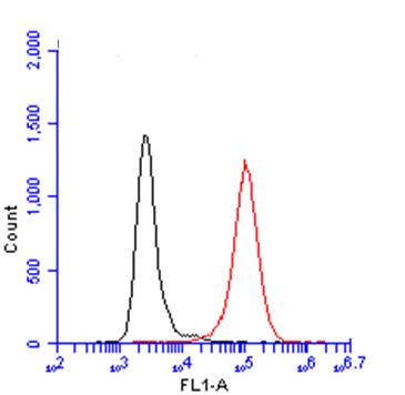
Iba1 antibody (GTX101495) detects Iba1 by flow cytometry analysis.
Sample: THP-1 cell.
Black: Unlabelled sample was used as a control.
Red: Iba1 antibody (GTX101495) dilution: 1:50.
GTX101495 WB Image
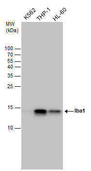
Iba1 antibody detects Iba1 protein by western blot analysis. Various whole cell extracts (30 ug) were separated by 15% SDS-PAGE, and the membrane was blotted with Iba1 antibody (GTX101495) diluted at a dilution of 1:5000. The HRP-conjugated anti-rabbit IgG antibody (GTX213110-01) was used to detect the primary antibody.
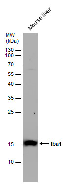
GTX101495 WB Image
Mouse tissue extract (50 ug) was separated by 15% SDS-PAGE, and the membrane was blotted with Iba1 antibody (GTX101495) diluted at 1:1000. The HRP-conjugated anti-rabbit IgG antibody (GTX213110-01) was used to detect the primary antibody.
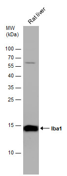
GTX101495 WB Image
Rat tissue extract (50 ug) was separated by 15% SDS-PAGE, and the membrane was blotted with Iba1 antibody (GTX101495) diluted at 1:1000. The HRP-conjugated anti-rabbit IgG antibody (GTX213110-01) was used to detect the primary antibody.
GTX101495 IHC-Fr Image
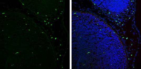
Iba1 antibody detects Iba1 protein expression at microglias by immunohistochemical analysis.
Sample: Frozen sectioned E13.5 Rat brain.
Green: Iba1 protein stained by Iba1 antibody (GTX101495) diluted at 1:250.
Red: beta Tubulin 3/ TUJ1, a mature neuron marker, stained by beta Tubulin 3/ TUJ1 antibody [GT11710] (GTX631836) diluted at 1:500.
Blue: Fluoroshield with DAPI (GTX30920).
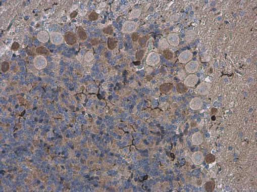
GTX101495 IHC-P Image
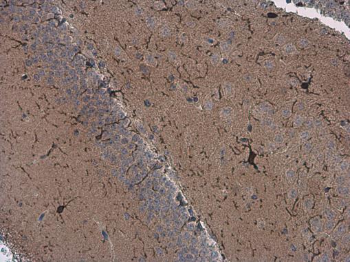
Iba1 antibody detects Iba1 protein at microglia in mouse brain by immunohistochemical analysis.
Sample: Paraffin-embedded mouse brain.
Iba1 antibody (GTX101495) diluted at 1:500.
GTX101495 IHC-P Image
Iba1 antibody detects Iba1 protein at microglia in rat brain by immunohistochemical analysis.
Sample: Paraffin-embedded rat brain.
Iba1 antibody (GTX101495) diluted at 1:500.
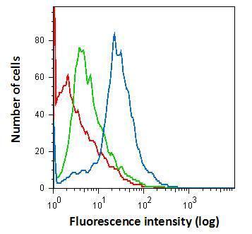
GTX101495 FACS Image
Flow cytometry on primary murine microglia cells, staining with Iba1 (GTX101495) antibody using 1.0 ug per 4Å~105 cells. GTX101495 (blue), Rabbit IgG (green) ,Unstained (red).