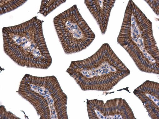
GTX101435 IHC-P Image
beta Catenin antibody [N1N2-2], N-term detects beta Catenin protein at membrane on mouse urinary bladder by immunohistochemical analysis.
Sample: Paraffin-embedded mouse urinary bladder.
beta Catenin antibody [N1N2-2], N-term (GTX101435) diluted at 1:500.
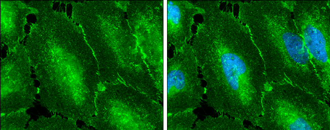
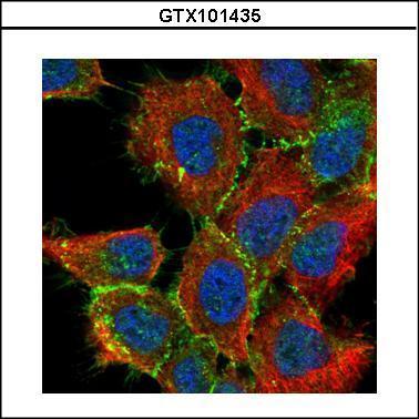
GTX101435 ICC/IF Image
beta Catenin antibody [N1N2-2], N-term detects beta Catenin protein at cell membrane by immunofluorescent analysis.
Sample: HeLa cells were fixed in 4% paraformaldehyde at RT for 15 min.
Green: beta Catenin protein stained by beta Catenin antibody [N1N2-2], N-term (GTX101435) diluted at 1:500.
Blue: Hoechst 33342 staining.
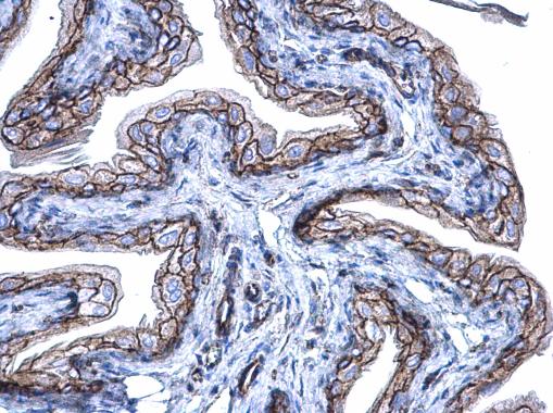
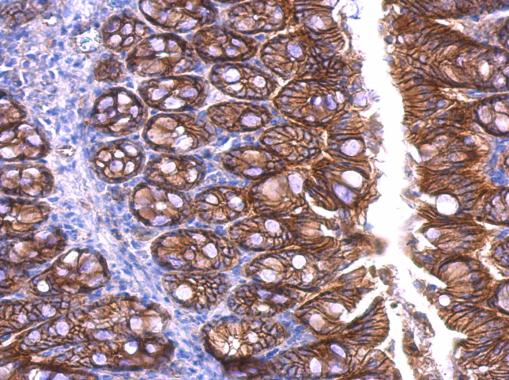
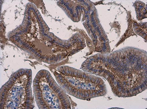
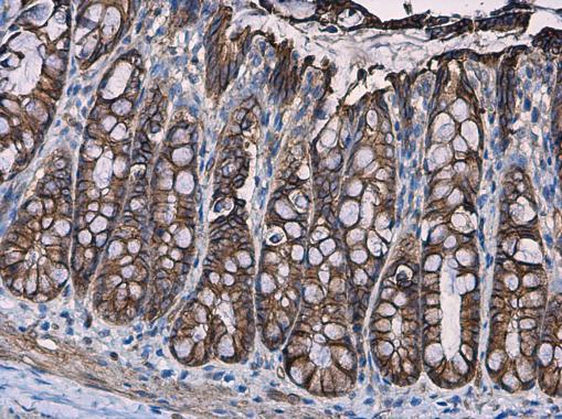
GTX101435 IHC-P Image
beta Catenin antibody [N1N2-2], N-term detects beta Catenin protein at cell membrane and cytoplasm in mouse duodenum by immunohistochemical analysis.
Sample: Paraffin-embedded mouse duodenum.
beta Catenin antibody [N1N2-2], N-term (GTX101435) diluted at 1:500.
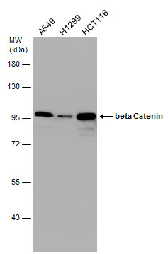
GTX101435 WB Image
Various whole cell extracts (30 ug) were separated by 7.5% SDS-PAGE, and the membrane was blotted with beta Catenin antibody [N1N2-2], N-term (GTX101435) diluted at 1:10000.
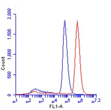
GTX101435 FACS Image
beta Catenin antibody [N1N2-2], N-term (GTX101435) detects CTNNB1 protein by flow cytometry analysis.
Sample: HeLa cell.
Black: Unlabelled sample was used as a control.
Red: beta Catenin antibody [N1N2-2], N-term (GTX101435) dilution: 1:50.
Acquisition of 20,000 events were collected using a Dylight 488-conjugated secondary antibody for FACS analysis.
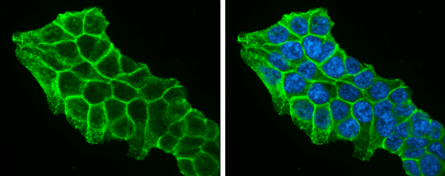
GTX101435 ICC/IF Image
beta Catenin antibody [N1N2-2], N-term detects beta Catenin protein at cell membrane by immunofluorescent analysis.
Sample: HCT 116 cells were fixed in 4% paraformaldehyde at RT for 15 min.
Green: beta Catenin protein stained by beta Catenin antibody [N1N2-2], N-term (GTX101435) diluted at 1:500.
Blue: Hoechst 33342 staining.
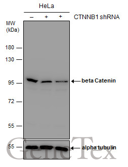
GTX101435 WB Image
Non-transfected (?) and transfected (+) HeLa whole cell extracts (30 ug) were separated by 7.5% SDS-PAGE, and the membrane was blotted with beta Catenin antibody [N1N2-2], N-term (GTX101435) diluted at 1:1000. The HRP-conjugated anti-rabbit IgG antibody (GTX213110-01) was used to detect the primary antibody.
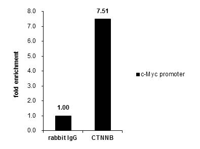
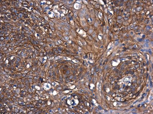
GTX101435 ChIP assay Image
Cross-linked ChIP was performed with HCT116 chromatin extract and 5 ug of either control rabbit IgG or anti-beta Catenin antibody. The precipitated DNA was detected by PCR with primer set targeting to c-Myc promoter.
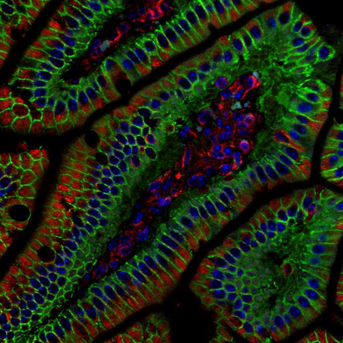
GTX101435 IHC-P Image
beta Catenin antibody [N1N2-2] detects beta Catenin protein at cell membrane in mouse colon by immunohistochemical analysis.
Sample: Paraffin-embedded mouse colon.
Green: beta Catenin antibody [N1N2-2] (GTX101435) diluted at 1:500.
Red: alpha Tubulin antibody [GT114] (GTX628802) diluted at 1:500.
Blue: Hoechst 33342 staining.
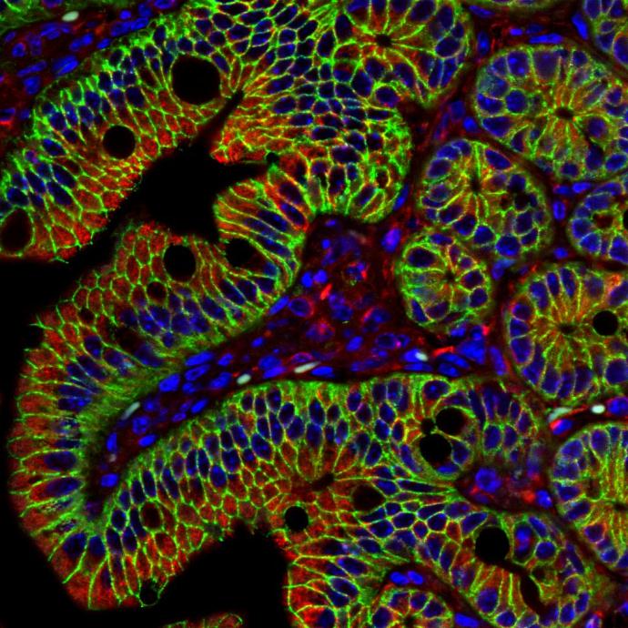
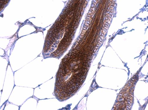
GTX101435 IHC-P Image
beta Catenin antibody [N1N2-2], N-term detects beta Catenin protein at membrane on mouse skin by immunohistochemical analysis.
Sample: Paraffin-embedded mouse skin.
beta Catenin antibody [N1N2-2], N-term (GTX101435) dilution: 1:500.
GTX101435 IHC-P Image
beta Catenin antibody [N1N2-2], N-term detects beta Catenin protein at membrane on mouse colon by immunohistochemical analysis.
Sample: Paraffin-embedded mouse colon.
beta Catenin antibody [N1N2-2], N-term (GTX101435) dilution: 1:500.
GTX101435 IHC-P Image
beta Catenin antibody [N1N2-2], N-term detects beta Catenin protein at cell membrane and cytoplasm in human esophagus by immunohistochemical analysis.
Sample: Paraffin-embedded human esophagus.
beta Catenin antibody [N1N2-2], N-term (GTX101435) diluted at 1:500.
GTX101435 IHC-P Image
beta Catenin antibody [N1N2-2], N-term detects beta Catenin protein at cell membrane and cytoplasm in human cervix by immunohistochemical analysis.
Sample: Paraffin-embedded human cervix.
beta Catenin antibody [N1N2-2], N-term (GTX101435) diluted at 1:500.
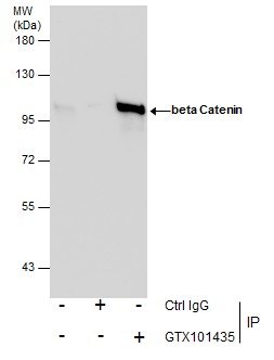
GTX101435 IP Image
Immunoprecipitation of beta Catenin protein from HeLa whole cell extracts using 5 ug of beta Catenin antibody (GTX101435).
Western blot analysis was performed using beta Catenin antibody (GTX101435).
EasyBlot anti-Rabbit IgG (GTX221666-01) was used as a secondary reagent.
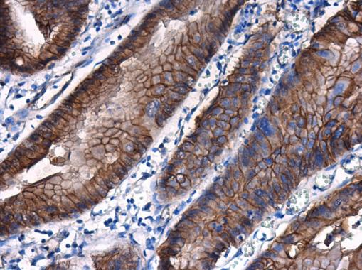
GTX101435 IHC-P Image
beta Catenin antibody [N1N2-2], N-term detects beta Catenin protein at cell membrane and cytoplasm in mouse duodenum by immunohistochemical analysis.
Sample: Paraffin-embedded mouse duodenum.
beta Catenin antibody [N1N2-2], N-term (GTX101435) diluted at 1:500.
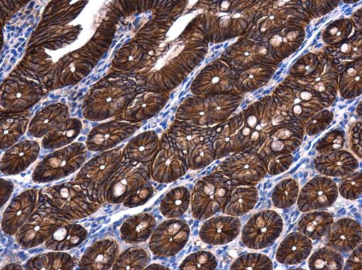
GTX101435 IHC-P Image
beta Catenin antibody [N1N2-2], N-term detects beta Catenin protein at cell membrane and cytoplasm in rat colon by immunohistochemical analysis.
Sample: Paraffin-embedded rat colon.
beta Catenin antibody [N1N2-2], N-term (GTX101435) diluted at 1:500.

GTX101435 IHC-P Image
beta Catenin antibody [N1N2-2], N-term detects beta Catenin protein at cell membrane and cytoplasm in rat duodenum by immunohistochemical analysis.
Sample: Paraffin-embedded rat duodenum.
beta Catenin antibody [N1N2-2], N-term (GTX101435) diluted at 1:500.
GTX101435 IHC-P Image
beta Catenin antibody [N1N2-2], N-term detects beta Catenin protein at cell membrane and cytoplasm in mouse intestine by immunohistochemical analysis.
Sample: Paraffin-embedded mouse intestine.
beta Catenin antibody [N1N2-2], N-term (GTX101435) diluted at 1:500.
GTX101435 ICC/IF Image
Confocal immunofluorescence analysis (Olympus FV10i) of paraformaldehyde-fixed A431, using beta Catenin(GTX101435) antibody (Green) at 1:200 dilution. Alpha-tubulin filaments were labeled with GTX11304 (Red) at 1:2000.