GTX101142 ICC/IF Image
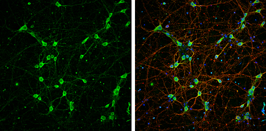
NF-L antibody detects NF-L protein at cytoplasm by immunofluorescent analysis.
Sample: DIV9 rat E18 primary cortical neurons were fixed in 4% paraformaldehyde at RT for 15 min.
Green: NF-L protein stained by NF-L antibody (GTX101142) diluted at 1:500.
Red: beta Tubulin 3/ Tuj1, stained by beta Tubulin 3/ Tuj1 antibody [GT11710] (GTX631836) diluted at 1:500.
Blue: Fluoroshield with DAPI (GTX30920).
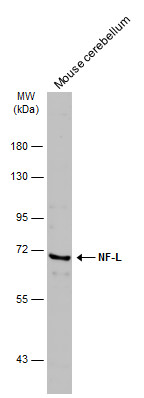
GTX101142 WB Image
Mouse tissue extract (5 ug) was separated by 7.5% SDS-PAGE, and the membrane was blotted with NF-L antibody (GTX101142) diluted at 1:500. The HRP-conjugated anti-rabbit IgG antibody (GTX213110-01) was used to detect the primary antibody.
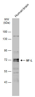
GTX101142 WB Image
Human brain (30 ug) was separated by 7.5% SDS-PAGE, and the membrane was blotted with NF-L antibody (GTX101142) diluted at 1:1000. The HRP-conjugated anti-rabbit IgG antibody (GTX213110-01) was used to detect the primary antibody.
GTX101142 IHC-Fr Image
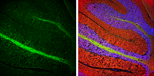
NF-L antibody detects NF-L protein expression by immunohistochemical analysis.
Sample: Frozen-sectioned adult mouse cerebellum.
Green: NF-L protein stained by NF-L antibody (GTX101142) diluted at 1:250.
Red: beta Tubulin 3/ TUJ1, stained by beta Tubulin 3/ TUJ1 antibody [GT11710] (GTX631836) diluted at 1:500.
Blue: Fluoroshield with DAPI (GTX30920).
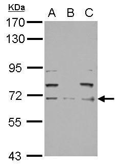
GTX101142 WB Image
Sample (30 ug of whole cell lysate)
A: PC-3
B: U87-MG
C: SK-N-SH
7.5% SDS PAGE
GTX101142 diluted at 1:10000
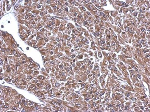
GTX101142 IHC-P Image
Immunohistochemical analysis of paraffin-embedded U87 xenograft, using 68kDa Neurofilament Light(GTX101142) antibody at 1:500 dilution.