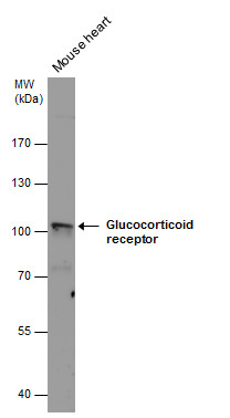
GTX101120 WB Image
Glucocorticoid receptor antibody detects Glucocorticoid receptor protein by western blot analysis. Mouse tissue extracts (50 ug) was separated by 7.5% SDS-PAGE, and the membrane was blotted with Glucocorticoid receptor antibody (GTX101120) diluted at 1:1000.
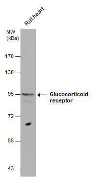
GTX101120 WB Image
Glucocorticoid receptor antibody detects Glucocorticoid receptor protein by western blot analysis. Rat tissue extracts (50 ug) was separated by 7.5% SDS-PAGE, and the membrane was blotted with Glucocorticoid receptor antibody (GTX101120) diluted at 1:1000.
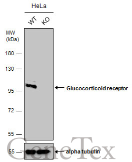
GTX101120 WB Image
Wild-type (WT) and Glucocorticoid receptor knockout (KO) HeLa cell extracts (30 ug) were separated by 7.5% SDS-PAGE, and the membrane was blotted with Glucocorticoid receptor antibody (GTX101120) diluted at 1:500. The HRP-conjugated anti-rabbit IgG antibody (GTX213110-01) was used to detect the primary antibody.
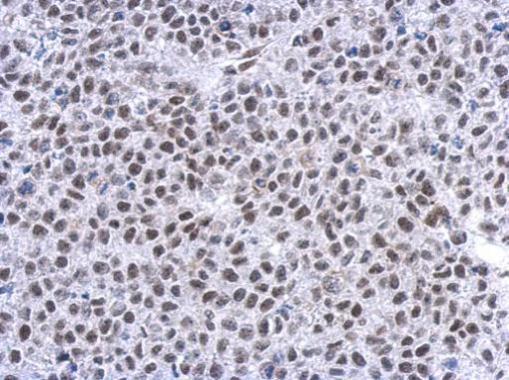
GTX101120 IHC-P Image
Immunohistochemical analysis of paraffin-embedded Hela xenograft, using Glucocorticoid receptor(GTX101120) antibody at 1:500 dilution.
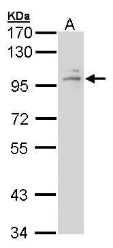
GTX101120 WB Image
Sample (30 ug of whole cell lysate)
A: A431 (GTX27909)
7.5% SDS PAGE
GTX101120 diluted at 1:1000
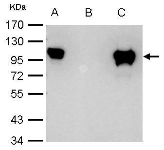
GTX101120 IP Image
Glucocorticoid receptor antibody immunoprecipitates Glucocorticoid receptor protein in IP experiments. IP Sample: 1000 ug HeLa whole cell lysate/extract A. 40 ug HeLa whole cell lysate/extract B. Control with 2.5 ug of preimmune rabbit IgG C. Immunoprecipitation of Glucocorticoid receptor protein by 2.5 ug of Glucocorticoid receptor antibody (GTX101120) 7.5% SDS-PAGE The immunoprecipitated Glucocorticoid receptor protein was detected by SCMH1 antibody (GTX101120) diluted at 1:1000. EasyBlot anti-rabbit IgG (GTX221666-01) was used as a secondary reagent.