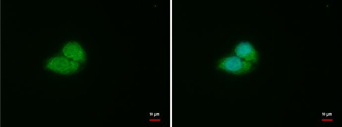
GTX101087 ICC/IF Image
VHL antibody detects VHL protein at cytoplasm and nucleus by immunofluorescent analysis.
Sample: HepG2 cells were fixed in 4% paraformaldehyde at RT for 15 min.
Green: VHL protein stained by VHL antibody (GTX101087) diluted at 1:500.
Blue: Hoechst 33342 staining.
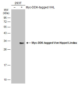
GTX101087 WB Image
Non-transfected (?) and transfected (+) 293T whole cell extracts (30 ug) were separated by 12% SDS-PAGE, and the membrane was blotted with Von Hippel Lindau antibody (GTX101087) diluted at 1:5000. The HRP-conjugated anti-rabbit IgG antibody (GTX213110-01) was used to detect the primary antibody.
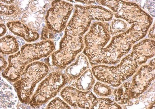
GTX101087 IHC-P Image
VHL antibody detects VHL protein at cytosol on mouse kidney by immunohistochemical analysis.
Sample: Paraffin-embedded mouse kidney.
VHL antibody (GTX101087) dilution: 1:500.
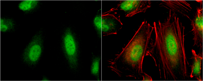
GTX101087 ICC/IF Image
VHL antibody detects VHL protein at cytoplasm and nucleus by immunofluorescent analysis.
Sample: HeLa cells were fixed in 4% paraformaldehyde at RT for 15 min.
Green: VHL protein stained by VHL antibody (GTX101087) diluted at 1:500.
Red: phalloidin, a cytoskeleton marker, stained by phalloidin (invitrogen, A12380) diluted at 1:200.
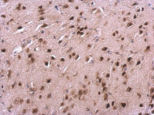
GTX101087 IHC-P Image
VHL antibody detects VHL protein at cytosol and nucleus on mouse fore brain by immunohistochemical analysis.
Sample: Paraffin-embedded mouse fore brain.
VHL antibody (GTX101087) dilution: 1:500.
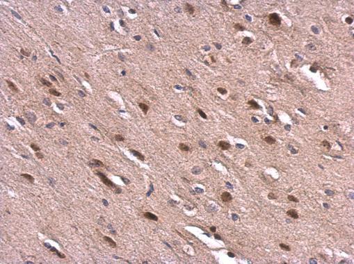
GTX101087 IHC-P Image
VHL antibody detects VHL protein at cytosol and nucleus on rat fore brain by immunohistochemical analysis.
Sample: Paraffin-embedded rat fore brain.
VHL antibody (GTX101087) dilution: 1:500.
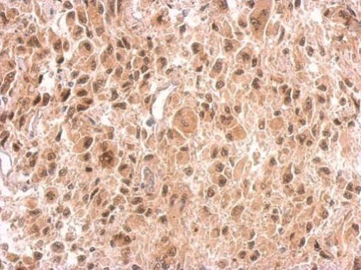
GTX101087 IHC-P Image
VHL antibody detects VHL protein at on U373 xenograft by immunohistochemical analysis.
Sample: Paraffin-embedded U373 xenograft.
VHL antibody (GTX101087) dilution: 1:500.