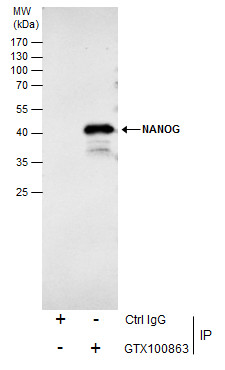
GTX100863 IP Image
Immunoprecipitation of NANOG protein from NT2D1 whole cell extracts using 5 ug of NANOG antibody [N3C3] (GTX100863).
Western blot analysis was performed using NANOG antibody [N3C3] (GTX100863).
EasyBlot anti-Rabbit IgG (GTX221666-01) was used as a secondary reagent.
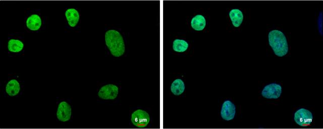
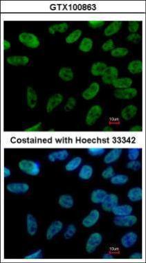
GTX100863 ICC/IF Image
NANOG antibody [N3C3] detects NANOG protein at nucleus by immunofluorescent analysis.
Sample: NT2D1 cells were fixed in 2% paraformaldehyde/culture medium at RT for 30 min.
Green: NANOG protein stained by NANOG antibody [N3C3] (GTX100863) diluted at 1:500.
Blue: Hoechst 33342 staining.
Scale bar = 6 um.
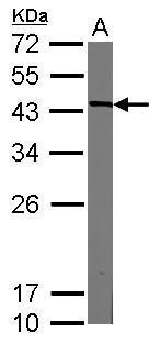
GTX100863 WB Image
Sample (30 ug of whole cell lysate)
A: HeLa nucleus
12% SDS PAGE
GTX100863 diluted at 1:3000
The HRP-conjugated anti-rabbit IgG antibody (GTX213110-01) was used to detect the primary antibody.
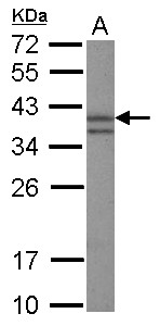
GTX100863 WB Image
Sample (30 ug of whole cell lysate)
A: NIH-3T3
10% SDS PAGE
GTX100863 diluted at 1:3000
The HRP-conjugated anti-rabbit IgG antibody (GTX213110-01) was used to detect the primary antibody.
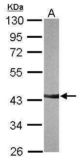
GTX100863 WB Image
Sample (20 ug of whole cell lysate)
A: mouse ESC
12% SDS PAGE
GTX100863 diluted at 1:3000
The HRP-conjugated anti-rabbit IgG antibody (GTX213110-01) was used to detect the primary antibody.
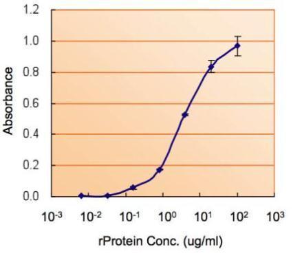
GTX100863 ELISA Image
ELISA detection of NANOG using GTX100863 for capture at a concentration of 5 ug/mL and GTX89079 for detection at a concentration of 5 ug/mL.
GTX100863 ICC/IF Image
Immunofluorescence analysis of paraformaldehyde-fixed Human ESC, using NANOG(GTX100863) antibody at 1:400 dilution.