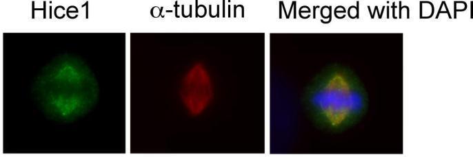
GTX100735 ICC/IF Image
Immunofluorescence analysis of human osteosarcoma cell line U2OS, using HICE1(GTX100735) antibody at 1:50-1:200 dilution.
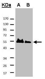
GTX100735 WB Image
Sample
A: His-Hice1 (2x)
B: His-Hice1 (1x)
7.5% SDS PAGE
GTX100735 diluted at 1:500
The HRP-conjugated anti-rabbit IgG antibody (GTX213110-01) was used to detect the primary antibody.
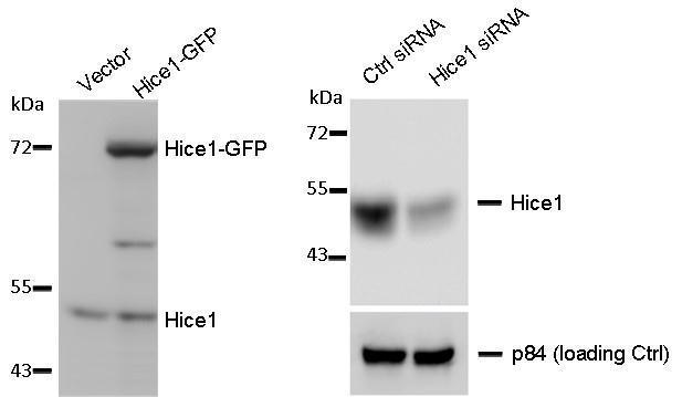
GTX100735 WB Image
WB to detect cellular Hice1 and Hice1-GFP expressed in human osteosarcoma U2OS cells (left image), and Hice1 upon siRNA treatment (right image), using GTX100735 at 1:1000 dilution. Nuclear matrix protein p84 is a loading control, blotted with p84 antibody (clone 5E10) GTX70220 at 1:5000 dilution. The HRP-conjugated anti-rabbit IgG antibody (GTX213110-01) was used to detect the primary antibody.
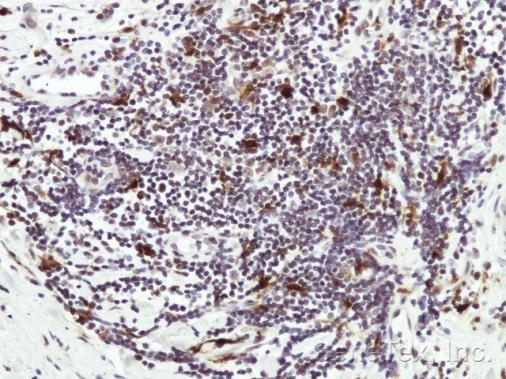
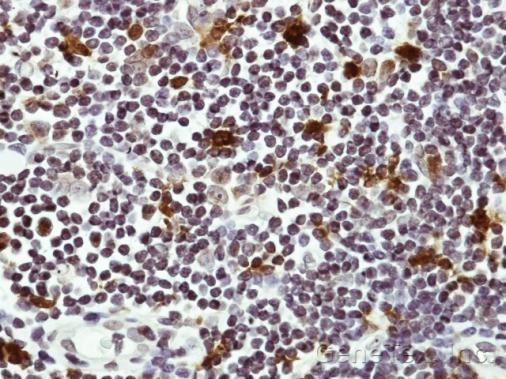
GTX100735 IHC-P Image
Immunohistochemical analysis of paraffin-embedded Human lymph tissue, using HICE1 (GTX100735) antibody at 1:50 dilution.
GTX100735 IHC-P Image
Immunohistochemical analysis of paraffin-embedded Human lymph tissue, using HICE1 (GTX100735) antibody at 1:50 dilution.
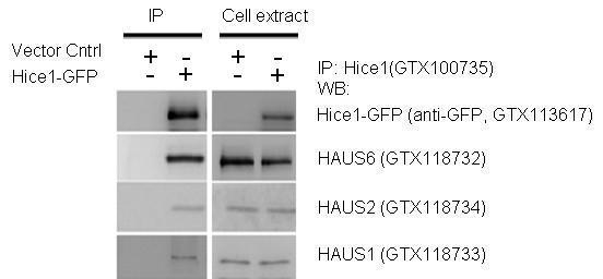
GTX100735 IP Image
IP-WB assay to show that Hice1 (GTX113617) co-immunoprecipitated with other Augmin components HAUS6 (GTX118732), HAUS2 (GTX118734) and HAUS1 (GTX118733) in U2OS cells.