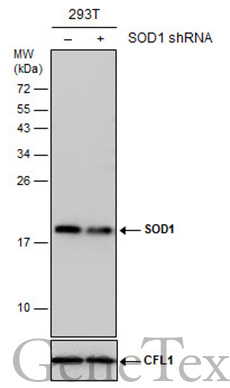
GTX100659 WB Image
Non-transfected (?) and transfected (+) 293T whole cell extracts (30 ug) were separated by 15% SDS-PAGE, and the membrane was blotted with SOD1 antibody (GTX100659) diluted at 1:5000. The HRP-conjugated anti-rabbit IgG antibody (GTX213110-01) was used to detect the primary antibody.
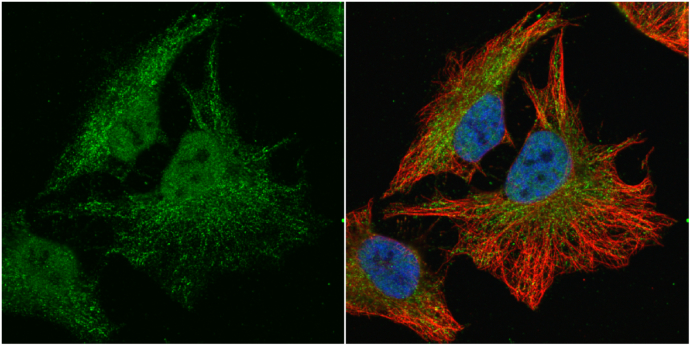
GTX100659 ICC/IF Image
SOD1 antibody detects SOD1 protein at cytoplasm and nucleus by immunofluorescent analysis.
Sample: HeLa cells were fixed in 4% paraformaldehyde at RT for 15 min.
Green: SOD1 protein stained by SOD1 antibody (GTX100659) diluted at 1:100.
Red: alpha Tubulin, a cytoskeleton marker, stained by alpha Tubulin antibody [GT114] (GTX628802) diluted at 1:1000.
Blue: Hoechst 33342 staining.
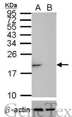
GTX100659 WB Image
Western blot analysis of SOD1 (GTX100659, upper panel) and beta-actin (GTX109639, lower panel)?
Sample (30 ug of whole cell lysate)?
A: HeLa mock control?
B: HeLa transfected shSOD1
15% SDS PAGE?
GTX100659 diluted at 1:500
The HRP-conjugated anti-rabbit IgG antibody (GTX213110-01) was used to detect the primary antibody.
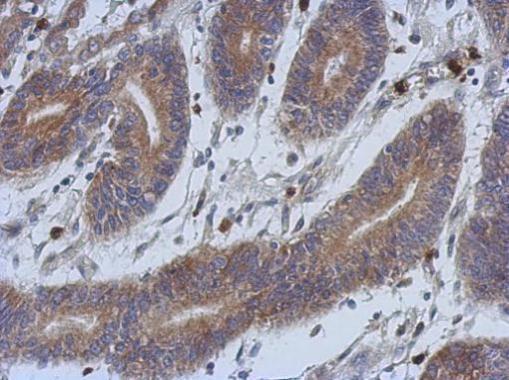
GTX100659 IHC-P Image
Immunohistochemical analysis of paraffin-embedded human colon carcinoma, using SOD1(GTX100659) antibody at 1:500 dilution.
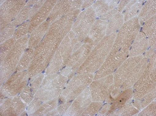
GTX100659 IHC-P Image
Immunohistochemical analysis of paraffin-embedded mouse muscle, using SOD1(GTX100659) antibody at 1:500 dilution.
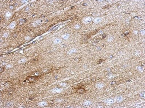
GTX100659 IHC-P Image
Immunohistochemical analysis of paraffin-embedded rat brain, using SOD1(GTX100659) antibody at 1:500 dilution.
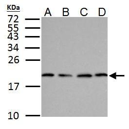
GTX100659 WB Image
SOD1 antibody detects SOD1 protein by western blot analysis.
A.30 ug NT2D1 whole cell lysate/extract
B.30 ug PC-3 whole cell lysate/extract
C.30 ug U87-MG whole cell lysate/extract
D.30 ug SK-N-SH whole cell lysate/extract
15% SDS-PAGE
SOD1 antibody (GTX100659) dilution: 1:500
The HRP-conjugated anti-rabbit IgG antibody (GTX213110-01) was used to detect the primary antibody.
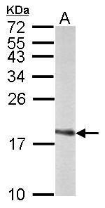
GTX100659 WB Image
Sample (50 ug of whole cell lysate)
A: Rat brain
15% SDS PAGE
GTX100659 diluted at 1:1000
The HRP-conjugated anti-rabbit IgG antibody (GTX213110-01) was used to detect the primary antibody.
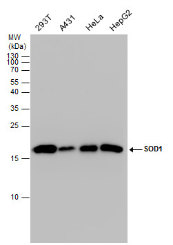
GTX100659 WB Image
SOD1 antibody detects SOD1 protein by western blot analysis. Various whole cell extracts (30 ug) were separated by 15% SDS-PAGE, and the membrane was blotted with SOD1 antibody (GTX100659) diluted by 1:1000. The HRP-conjugated anti-rabbit IgG antibody (GTX213110-01) was used to detect the primary antibody.