
GTX100579 IHC-P Image
TARDBP antibody detects TARDBP protein at nucleus in mouse brain by immunohistochemical analysis.
Sample: Paraffin-embedded mouse brain.
TARDBP antibody (GTX100579) diluted at 1:500.
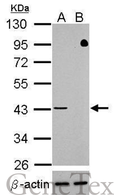
GTX100579 WB Image
Western blot analysis of TDP-43 (GTX100579, upper panel) and beta-actin (GTX109639, lower panel)?
Sample (30 ug of whole cell lysate)
? A: HeLa mock control?
B: HeLa transfected shTDP-43
10% SDS PAGE?
GTX100579 diluted at 1:500
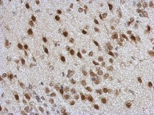
GTX100579 IHC-P Image
Immunohistochemical analysis of paraffin-embedded rat brain, using TARDBP(GTX100579) antibody at 1:500 dilution.
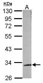
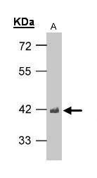
GTX100579 WB Image
Sample (50 ug of whole cell lysate)
A: mouse brain
10% SDS PAGE
GTX100579 diluted at 1:500
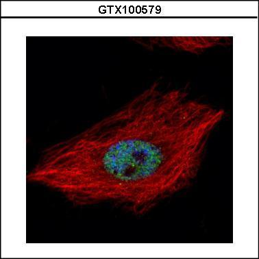
GTX100579 ICC/IF Image
Confocal immunofluorescence analysis (Olympus FV10i) of paraformaldehyde-fixed HeLa, using TARDBP(GTX100579) antibody (Green) at 1:500 dilution. Alpha-tubulin filaments were labeled with GTX11304 (Red) at 1:2000.
GTX100579 WB Image
Sample(30 ug of whole cell lysate)
A:H1299
10% SDS PAGE
GTX100579 diluted at 1:1000