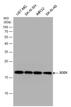
GTX100554 WB Image
SOD1 antibody detects SOD1 protein by western blot analysis. Various whole cell extracts (30 ug) were separated by 15% SDS-PAGE, and the membrane was blotted with SOD1 antibody (GTX100554) diluted at a dilution of 1:1000. The HRP-conjugated anti-rabbit IgG antibody (GTX213110-01) was used to detect the primary antibody.
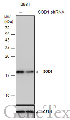
GTX100554 WB Image
Non-transfected (?) and transfected (+) 293T whole cell extracts (30 ug) were separated by 15% SDS-PAGE, and the membrane was blotted with SOD1 antibody (GTX100554) diluted at 1:5000. The HRP-conjugated anti-rabbit IgG antibody (GTX213110-01) was used to detect the primary antibody.
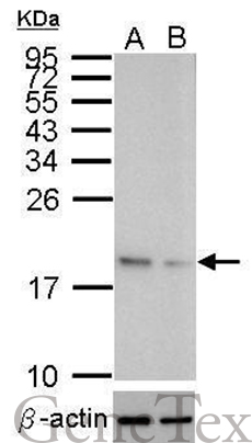
GTX100554 WB Image
Western blot analysis of SOD1 (GTX100554, upper panel) and beta-actin (GTX109639, lower panel)?
Sample (10 ug of whole cell lysate)?
A: HeLa mock control?
B: HeLa transfected shSOD1
15% SDS PAGE?
GTX100554 diluted at 1:500
The HRP-conjugated anti-rabbit IgG antibody (GTX213110-01) was used to detect the primary antibody.
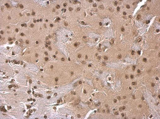
GTX100554 IHC-P Image
SOD1 antibody detects SOD1 protein at cytosol on mouse fore brain by immunohistochemical analysis.
Sample: Paraffin-embedded mouse fore brain.
SOD1 antibody (GTX100554) dilution: 1:500.
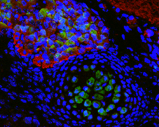
GTX100554 IHC-P Image
Immunofluorescence photomicrographs of paraffin-embedded mouse fetal brain.
Green: SOD1 antibody (GTX100554) diluted at 1:200. The signal was developed using goat anti-rabbit IgG antibody (Dylight488) (GTX213110-04).
Red: beta Tubulin 3/ TUJ1 antibody [GT11710] diluted at 1:100. The signal was developed using goat anti-mouse IgG antibody (Dylight594) (GTX213111-05).
Blue: Nuclear staining with Hoechst 33342.
GTX100554 WB Image
SOD1 antibody detects SOD1 protein by western blot analysis. Whole cell extracts (30 ug) was separated by 15% SDS-PAGE, and the membrane was blotted with SOD1 antibody (GTX100554) at a dilution of 1:1000. The HRP-conjugated anti-rabbit IgG antibody (GTX213110-01) was used to detect the primary antibody.

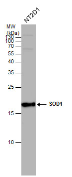
GTX100554 WB Image
Sample (50 ug of whole cell lysate)
A: mouse brain
15% SDS PAGE
GTX100554 diluted at 1:1000
The HRP-conjugated anti-rabbit IgG antibody (GTX213110-01) was used to detect the primary antibody.
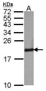
GTX100554 WB Image
Sample (30 ug of whole cell lysate)
A: HeLa
15% SDS PAGE
Superoxide Dismutase 1 antibody
GTX100554 diluted at 1:1000
The HRP-conjugated anti-rabbit IgG antibody (GTX213110-01) was used to detect the primary antibody.
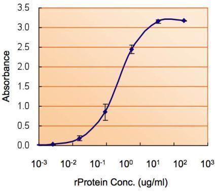
GTX100554 ELISA Image
ELISA detection of SOD1 using GTX100554 for capture at a concentration of 5 ug/mL and GTX89049 for detection at a concentration of 1.5 ug/mL.

GTX100554 WB Image
Sample (50 ug of whole cell lysate)
A: Rat brain
15% SDS PAGE
GTX100554 diluted at 1:1000
The HRP-conjugated anti-rabbit IgG antibody (GTX213110-01) was used to detect the primary antibody.
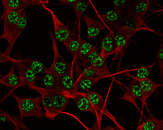
GTX100554 ICC/IF Image
SOD1 antibody detects SOD1 protein at nucleus by immunofluorescent analysis.
Sample: U-87 MG cells were fixed in 4% paraformaldehyde at RT for 15 min.
Green: SOD1 protein stained by SOD1 antibody (GTX100554) diluted at 1:500.
Red: beta Tubulin 3/ TUJ1 protein stained by beta Tubulin 3/ TUJ1 antibody (GTX631836) diluted at 1:200.