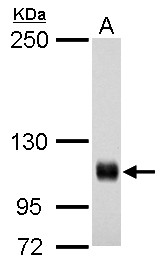
GTX100545 WB Image
Rb antibody detects Rb protein by western blot analysis.
A. 30 ug A431 whole cell lysate/extract
5% SDS-PAGE
Rb antibody (GTX100545) dilution: 1:500
The HRP-conjugated anti-rabbit IgG antibody (GTX213110-01) was used to detect the primary antibody.
GTX100545 IP Image
Rb antibody immunoprecipitates retinoblastoma 1 protein in IP experiments.
IP samples: HeLa whole cell extract
A. Control with 4 ug of preimmune Rabbit IgG
B. Immunoprecipitation of retinoblastoma 1 protein by 4 ug Rb antibody (GTX100545)
7.5 % SDS-PAGE
The immunoprecipitated retinoblastoma 1 protein was detected by Rb antibody (GTX100545) diluted at 1:500.
[EasyBlot anti-rabbit IgG (GTX221666-01) was used as a secondary reagent]
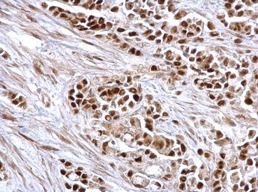
GTX100545 IHC-P Image
Rb antibody detects Rb protein at nucleus on human colon carcinoma by immunohistochemical analysis.
Sample: Paraffin-embedded human colon carcinoma.
Rb antibody (GTX100545) dilution: 1:500.
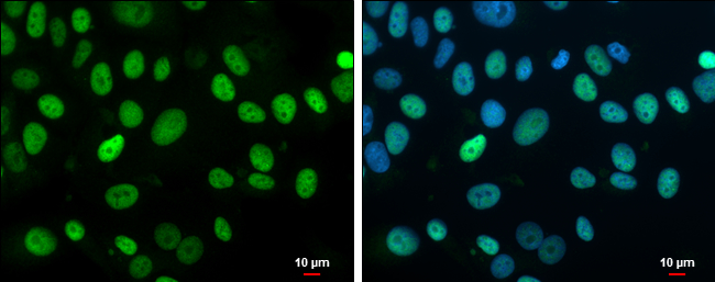
GTX100545 ICC/IF Image
Rb antibody detects Rb protein at nucleus by immunofluorescent analysis.
Sample: MCF7 cells were fixed in 4% paraformaldehyde at RT for 15 min.
Green: Rb protein stained by Rb antibody (GTX100545) diluted at 1:500.
Blue: Hoechst 33342 staining.
Scale bar = 10 um.
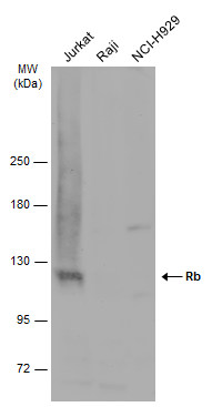
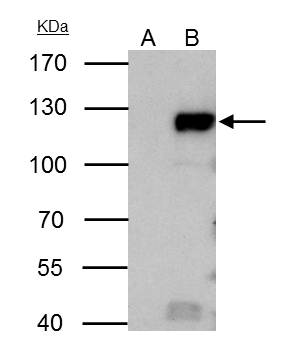
GTX100545 WB Image
Various whole cell extracts (30 ug) were separated by 5% SDS-PAGE, and the membrane was blotted with Rb antibody (GTX100545) diluted at 1:1000. The HRP-conjugated anti-rabbit IgG antibody (GTX213110-01) was used to detect the primary antibody.