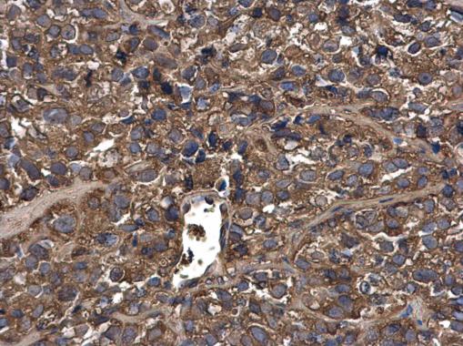
GTX100459 IHC-P Image
MUC1 antibody detects MUC1 protein at cytoplasm in human cervical cancer by immunohistochemical analysis.
Sample: Paraffin-embedded human cervical cancer.
MUC1 antibody (GTX100459) diluted at 1:500.
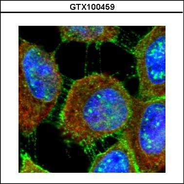
GTX100459 ICC/IF Image
Confocal immunofluorescence analysis (Olympus FV10i) of methanol-fixed A431, using MUC1(GTX100459) antibody (Green) at 1:500 dilution. Alpha-tubulin filaments were labeled with GTX11304 (Red) at 1:500.
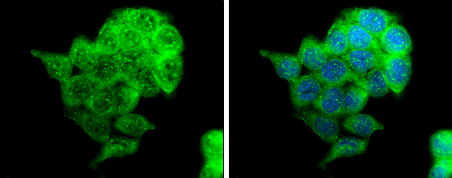
GTX100459 ICC/IF Image
MUC1 antibody detects MUC1 protein at cytoplasm by immunofluorescent analysis.
Sample: HCT116 cells were fixed in ice-cold MeOH for 5 min.
Green: MUC1 protein stained by MUC1 antibody (GTX100459) diluted at 1:500.
Blue: Hoechst 33342 staining.
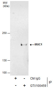
GTX100459 IP Image
Immunoprecipitation of MUC1 protein from HCT-116 whole cell extracts using 5 ug of MUC1 antibody (GTX100459).
Western blot analysis was performed using MUC1 antibody (GTX100459).
EasyBlot anti-Rabbit IgG (GTX221666-01) was used as a secondary reagent.
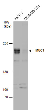
GTX100459 WB Image
Various whole cell extracts (30 ug) were separated by 5% SDS-PAGE, and the membrane was blotted with MUC1 antibody (GTX100459) diluted at 1:1000.