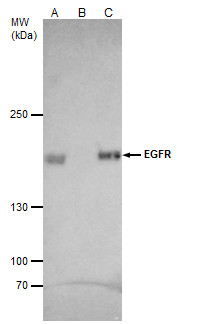
GTX100448 IP Image
EGFR antibody [C2C3], C-term immunoprecipitates EGFR protein in IP experiments.
IP samples: A431 whole cell extract
A. 40 ug A431 whole cell extract
B. Control with 4 ug of preimmune Rabbit IgG
C. Immunoprecipitation of EGFR protein by 4 ug EGFR antibody [C2C3], C-term (GTX100448)
5 % SDS-PAGE
The immunoprecipitated EGFR protein was detected by EGFR antibody [C2C3], C-term (GTX100448) diluted at 1:1000.
[EasyBlot anti-rabbit IgG (GTX221666-01) was used as a secondary reagent]
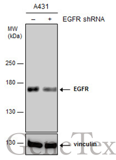
GTX100448 WB Image
Non-transfected (?) and transfected (+) A431 whole cell extracts (15 ug) were separated by 5% SDS-PAGE, and the membrane was blotted with EGFR antibody [C2C3], C-term (GTX100448) diluted at 1:6000. The HRP-conjugated anti-rabbit IgG antibody (GTX213110-01) was used to detect the primary antibody.
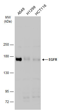
GTX100448 WB Image
Various whole cell extracts (30 ug) were separated by 5% SDS-PAGE, and the membrane was blotted with EGFR antibody [C2C3], C-term (GTX100448) diluted at 1:500. The HRP-conjugated anti-rabbit IgG antibody (GTX213110-01) was used to detect the primary antibody.
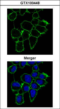
GTX100448 ICC/IF Image
Confocal immunofluorescence analysis (Olympus FV10i) of methanol-fixed A431, using EGFR(GTX100448) antibody (Green) at 1:1000 dilution.
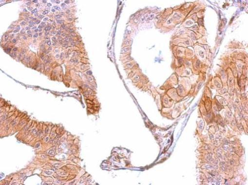
GTX100448 IHC-P Image
EGFR antibody [C2C3], C-term detects EGFR protein at membrane on human gastric cancer by immunohistochemical analysis.
Sample: Paraffin-embedded gastric cancer .
EGFR antibody [C2C3], C-term (GTX100448) dilution: 1:500.
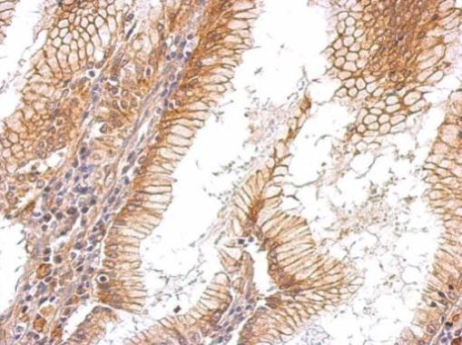
GTX100448 IHC-P Image
EGFR antibody [C2C3], C-term detects EGFR protein at membrane on human gastric cancer by immunohistochemical analysis.
Sample: Paraffin-embedded gastric cancer.
EGFR antibody [C2C3], C-term (GTX100448) dilution: 1:500.