GTX100446 IHC-Fr Image
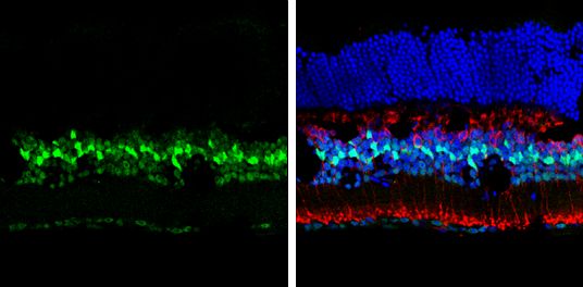
p27 Kip1 antibody detects p27 Kip1 protein at nucleus by immunohistochemical analysis.
Sample: Frozen sectioned adult mouse retina.
Green: p27 Kip1 protein stained by p27 Kip1 antibody (GTX100446) diluted at 1:250.
Red: Protein kinase C alpha staining.
Blue: Fluoroshield with DAPI (GTX30920).
GTX100446 WB Image
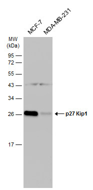
Various whole cell extracts (30 ug) were separated by 12% SDS-PAGE, and the membrane was blotted with p27 Kip1 antibody (GTX100446) diluted at 1:1000. The HRP-conjugated anti-rabbit IgG antibody (GTX213110-01) was used to detect the primary antibody.
GTX100446 IP Image
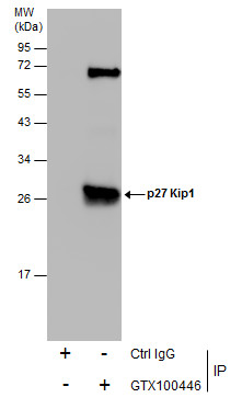
Immunoprecipitation of p27 Kip1 protein from MCF-7 whole cell extracts using 5 ug of p27 Kip1 antibody (GTX100446).
Western blot analysis was performed using p27 Kip1 antibody (GTX100446).
EasyBlot anti-Rabbit IgG (GTX221666-01) was used as a secondary reagent.
GTX100446 WB Image
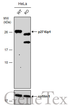
Wild-type (WT) and p27 Kip1 knockout (KO) HeLa cell extracts (30 ug) were separated by 12% SDS-PAGE, and the membrane was blotted with p27 Kip1 antibody (GTX100446) diluted at 1:500. The HRP-conjugated anti-rabbit IgG antibody (GTX213110-01) was used to detect the primary antibody.

GTX100446 IHC-P Image
Immunohistochemical staining of human ulcerative colitis tissue using anti-p27 Kip1 antibody (GTX100446).
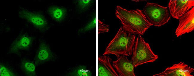
GTX100446 ICC/IF Image
p27 Kip1 antibody detects p27 Kip1 protein at cytoplasm and nucleus by immunofluorescent analysis.
Sample: HeLa cells were fixed in 4% paraformaldehyde at RT for 15 min.
Green: p27 Kip1 protein stained by p27 Kip1 antibody (GTX100446) diluted at 1:500.
Red: phalloidin, a cytoskeleton marker, diluted at 1:200.
Scale bar = 10 um.
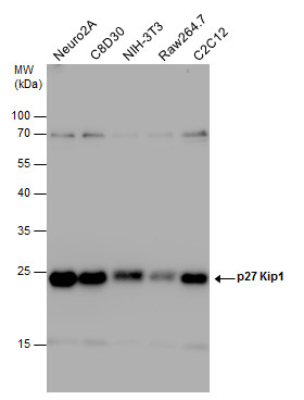
GTX100446 WB Image
p27 Kip1 antibody detects p27 Kip1 protein by Western blot analysis. Various whole cell extracts (30 ug) were separated by 12% SDS-PAGE, and the membrane was blotted with p27 Kip1 antibody (GTX100446) diluted at 1:1000.