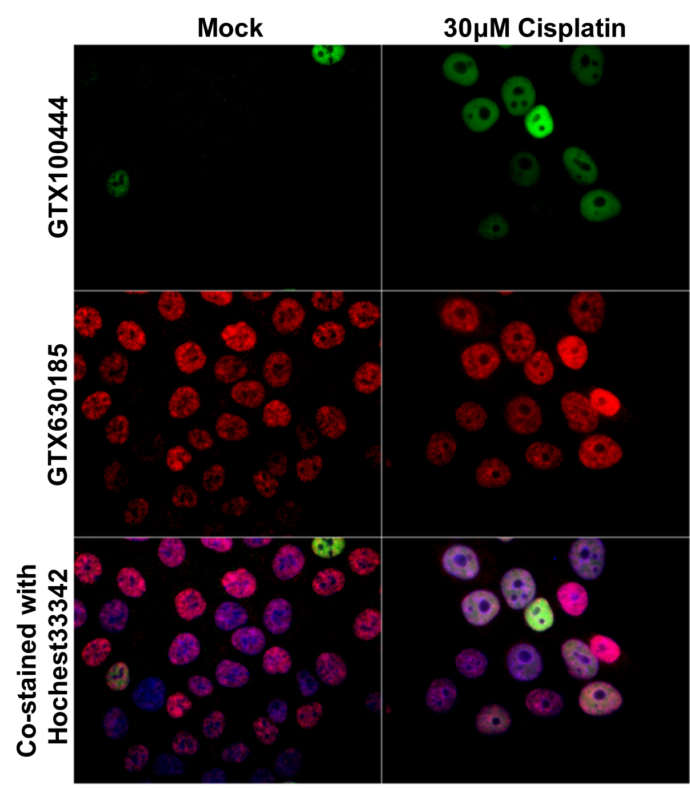
GTX100444 ICC/IF Image
p21 Cip1 antibody detects p21 Cip1 protein at nucleus by immunofluorescent analysis.
Samples: HCT116 cells mock (left) and treated with 30 uM Cisplatin for 24 hrs (right) were fixed in 4% paraformaldehyde at RT for 15 min.
Green: p21 Cip1 protein stained by p21 Cip1 antibody (GTX100444) diluted at 1:1000.
Red: Histone H3S10ph (phospho Ser10), a nucleus marker, stained by Histone H3S10ph (phospho Ser10) antibody [GT921] (GTX630185) diluted at 1:500.
Blue: Hoechst 33342 staining.
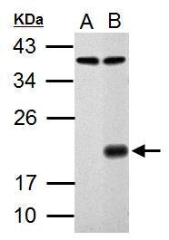
GTX100444 WB Image
Sample (30 ug of whole cell lysate)
A: HCT116
B: HCT116 treated with 30 uM cisplatin for 24 hr
12% SDS PAGE
GTX100444 diluted at 1:1000
The HRP-conjugated anti-rabbit IgG antibody (GTX213110-01) was used to detect the primary antibody.